

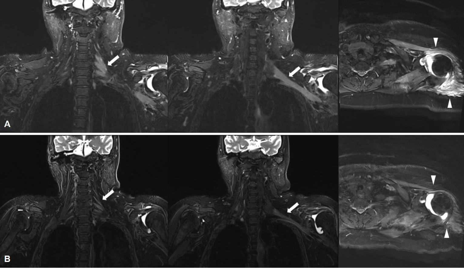
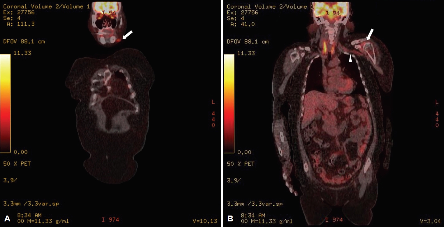
67세 여자가 2달 전 발생한 왼쪽 팔의 근력저하로 방문하였다. 신경전도검사에서 왼쪽 정중신경, 노신경, 근피부신경에서 이상이 관찰되고 침근전도검사에서 집게폄근, 원엎침근, 위팔두갈래근 등에 비정상 자발전위가 관찰되어, 팔신경얼기병으로 진단하였다. 어깨 자기공명영상에서 왼쪽 팔신경얼기의 비대신경병이 의심되었다(Fig. 1-A). 입원 후 왼쪽 턱의 발진 부위와 왼쪽 겨드랑이에서 실시한 생검에서 광범위큰B세포림프종을 확인하였다. 양전자방출단층촬영(positron emission tomography)에서 왼쪽 턱, 겨드랑이, 왼쪽 팔신경얼기에서 대사 증가가 관찰되었다(Fig. 2). 항암화학 치료 이후 실시한 자기공명영상에서 비대신경병의 호전이 있었다(Fig. 1-B).
| J Korean Neurol Assoc > Volume 39(2); 2021 > Article |

|
REFERENCES
1. Khadilkar SV, Yadav RS, Soni G. A practical approach to enlargement of nerves, plexuses and roots. Pract Neurol 2015;15:105-115.


2. Bourque PR, Warman Chardon J, Bryanton M, Toupin M, Burns BF, Torres C. Neurolymphomatosis of the brachial plexus and its branches: case series and literature review. Can J Neurol Sci 2018;45:137-143.
Figure 1.
Coronal and axial MRI in the pre-treatment (A) and post-treatment stage (B). Coronal fat-suppressed T2-weighted MRI images show marked thickening and increased signal intensity along the left brachial plexus (arrows) and atrophic changes in the shoulder muscles (arrowheads) (A), and later improved in the affected area (B). MRI; magnetic resonance imaging.
- TOOLS
 PDF Links
 PubReader
 ePub Link
 Full text via DOI
 Download Citation
 Print
Print


-
METRICS

-
- 0 Crossref
- 0 Scopus
- 1,110 View
- 42 Download
-

- Related articles
-
Neurofibromatosis Type 1 with Idiopathic Hypertrophic Pachymeningitis2022 May;40(2)
Neurosyphilis Presenting with Status Epilepticus2020 November;38(4)
ANCA-Associated Vasculitis Presenting with Hypertrophic Pachymeningitis2018 August;36(3)
Neurosyphilis Presenting with Unilateral Tonic Pupil2011 ;29(3)
Neurosarcoidosis Presenting as Spinal Nerve Root Pain of Trunk2010 ;28(2)
- Editorial Office
-
(ZIP 03163) #1111, Daeil Bldg, 12, Insadong-gil, Jongno-gu, Seoul, Korea
Tel: +82-2-737-6530 Fax: +82-2-737-6531 E-mail: jkna@neuro.or.kr
Copyright © 2024 by Korean Neurological Association.

