새롭게 발견된 후교통동맥류에 의한 통증눈돌림신경마비

Painful Oculomotor Nerve Palsy Caused by a Newly Detected Posterior Communicating Artery Aneurysm

Article information

J Korean Neurol Assoc. 2026;44(3):244-245
Publication date (electronic) : August 1, 2026
doi : http://dx.doi.org/10.17340/jkna.2026.0014
Health Service Section, Republic of Korea Army, Seoul, Korea
aDepartment of Neurology, VHS Medical Center, Seoul, Korea
정원철, 이슬a, 김정섭a, 윤보혜a, 김승민,a
대한민국 국군 의무대
a중앙보훈병원 신경과
Address for correspondence Seung Min Kim, MD Department of Neurology, VHS Medical Center, 53 Jinhwangdo-ro 61-gil, Gangdonggu, Seoul 05368, Korea Tel: +82-2-2225-1324 Fax: +82-2-2225-1327 E-mail: kh21762@hanmail.net
received : February 27, 2026 , rev-recd : March 26, 2026 , accepted : March 27, 2026 .

고혈압, 고지혈증, 대동맥판막치환술 병력이 있던 77세 남자가 갑작스러운 좌측 안와 통증과 안검하수, 동공부등을 동반한 완전눈돌림신경마비로 내원하였다. 동공 침범을 동반한 통증눈돌림신경마비는 후교통동맥류에 의한 압박성 병변을 우선적으로 고려해야 하는 증상이지만[1] 13개월 전 시행한 뇌자기공명혈관조영술에서는 두개 내 동맥류가 관찰되지 않았다. 그러나 추적 영상 검사에서 이전에는 보이지 않던 3 mm 크기의 좌측 후교통동맥류가 새롭게 확인되었다(Fig. 1). 즉각적인 코일색전술 후 안와 통증은 빠르게 호전되었고 눈돌림신경기능도 점진적으로 회복되었다(Fig. 2). 신생(de novo) 동맥류의 빠른 형성은 혈관벽 취약이나 국소 혈역학 스트레스와 관련될 수 있다[2]. 본 증례는 최근 영상이 정상이라 하더라도 고혈압, 고지혈증 등의 혈관 위험인자를 지닌 상황에서는 짧은 기간 안에 신생 동맥류가 형성될 수 있음을 반드시 고려해야 함을 시사한다[3].
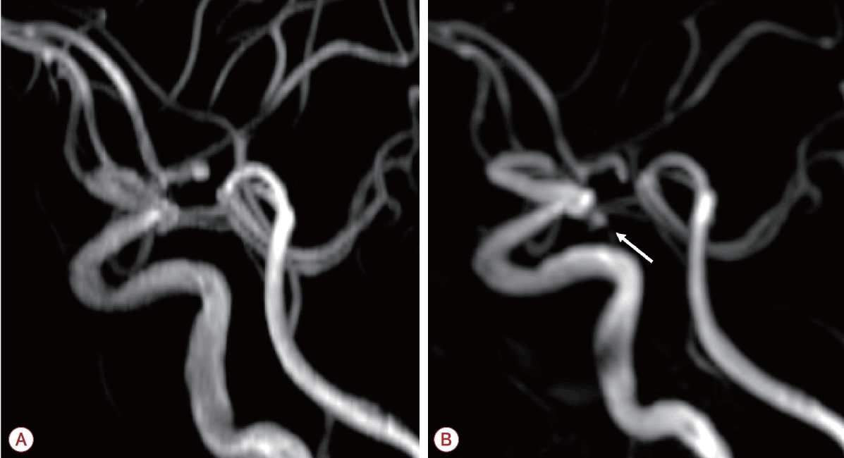
Figure 1.

(A) Intracranial MRA performed 13 months ago shows no significant aneurysmal dilatation. (B) Follow-up MRA reveals a de novo 3 mm aneurysm (white arrow) at the left posterior communicating artery. MRA; magnetic resonance angiography.
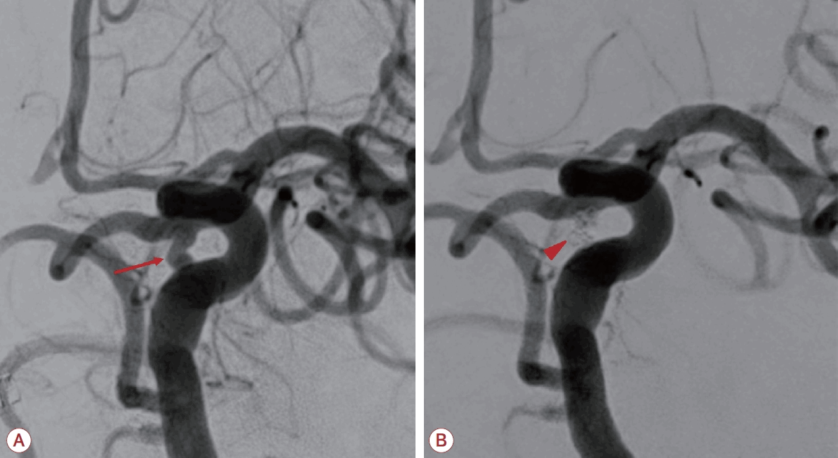
Figure 2.

(A) Transfemoral cerebral angiography demonstrates a small de novo posterior communicating artery aneurysm (red arrow). (B) Post-treatment angiography demonstrates complete coil embolizations of the aneurysm with no residual filling (red arrowhead).

References

1. Keane JR. Third nerve palsy: analysis of 1400 personally-examined inpatients. Can J Neurol Sci 2010;37:662–670.
2. Ferns SP, Sprengers ME, van Rooij WJ, van den Berg R, Velthuis BK, de Kort GA, et al. De novo aneurysm formation and growth of untreated aneurysms: a 5-year MRA follow-up in a large cohort of patients with coiled aneurysms and review of the literature. Stroke 2011;42:313–318.
3. da Costa MDS, Lima JVF, Zanini MA, Filho PTH, Naufal RFF, Reys L, et al. Risks for oculomotor nerve palsy and time to recovery after surgical clipping of posterior communicating artery aneurysms: a multicenter retrospective cohort study. Neurosurgery 2023;92:1192–1198.

Article information Continued

Figure 1.

(A) Intracranial MRA performed 13 months ago shows no significant aneurysmal dilatation. (B) Follow-up MRA reveals a de novo 3 mm aneurysm (white arrow) at the left posterior communicating artery. MRA; magnetic resonance angiography.

Figure 2.

(A) Transfemoral cerebral angiography demonstrates a small de novo posterior communicating artery aneurysm (red arrow). (B) Post-treatment angiography demonstrates complete coil embolizations of the aneurysm with no residual filling (red arrowhead).