

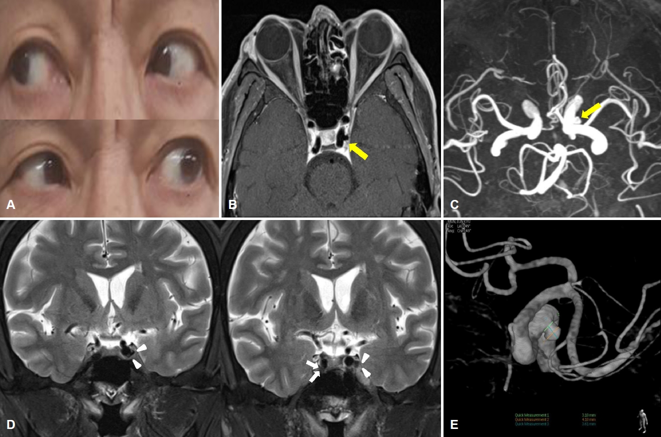
53세 남자가 5일 전에 발생한 복시로 내원하였다. 기저질환은 간경화가 있었고 만성 신부전증으로 혈액 투석 중이었다. 신경계 진찰에서 좌안의 외전에 제한이 있었고, 20 cm 이상 주시하였을 때 복시증상이 악화되어 좌안의 외전신경마비가 의심되었다(Fig. A). 뇌 자기공명영상(Fig. B, D) 및 뇌 자기공명혈관조영에서 비파열 내경동맥류가 보여(Fig. C) 뇌혈관조영술을 시행하였다. 해면정맥동 부위의 내경동맥에서 4×4 mm 크기의 목이 넓은 뇌동맥류를 확인하였으나(Fig. E) 시술의 적응증에 해당하지 않아 추가 약물 없이 외래에서 경과 관찰하고 있으며 복시는 계속 남아있는 상태이다. 해면정맥동 부위의 내경동맥류에 의한 외전신경마비는 3% 정도에서 나타나며, 증례 환자와 같이 단독 외전신경마비를 보인 경우는 드물다[1]. 외전신경은 해면정맥동내에서 내경동맥 덮개의 내측면에 위치하므로 내경동맥류의 기계적 압박이나 신경의 허혈에 의해 마비가 생길 수 있다[2]. 이전 보고에서는 10 mm 이상의 큰 동맥류에서 뇌신경마비를 보였으나, 본 증례는 4 mm의 동맥류에서 나타나 작은 내경동맥류도 단독 외전신경마비의 원인이 될 수 있음을 시사한다.
| J Korean Neurol Assoc > Volume 39(3); 2021 > Article |

|
Figure.
(A) Lateral gaze limitation in the left eye. (B) Orbit magnetic resonance imaging reveals a cavernous aneurysm (arrow) of the left internal carotid artery. (C) Brain magnetic resonance angiography shows a 4-mm aneurysm (arrow) in the cavernous portion of left internal carotid artery. (D) Brain magnetic resonance imaging (coronal view) shows the abducens nerve (white arrows) on the right. Abducens nerve is not visible on the left due to aneurysm (white arrowheads). (E) Transfemoral cerebral angiography reveals a saccular aneurysm of internal carotid artery.
- TOOLS
 PDF Links
 PubReader
 ePub Link
 Full text via DOI
 Download Citation
 Print
Print


-
METRICS

-
- 0 Crossref
- 0 Scopus
- 957 View
- 41 Download
-

- Related articles
-
Isolated Hypoglossal Nerve Palsy Caused by Dural Arteriovenous Fistula2016 November;34(4)
Isolated Bilateral Abducens Nerve Palsy Caused by Basilar Artery Dissecting Aneurysm2012 ;30(4)
Unilateral Abducens Nerve Palsy due to Bilateral Dural Carotid Cavernous Fistula2009 ;27(3)
Isolated Trigeminal Neuropathy Caused by Pontine Infarction2009 ;27(2)
- Editorial Office
-
(ZIP 03163) #1111, Daeil Bldg, 12, Insadong-gil, Jongno-gu, Seoul, Korea
Tel: +82-2-737-6530 Fax: +82-2-737-6531 E-mail: jkna@neuro.or.kr
Copyright © 2024 by Korean Neurological Association.

