

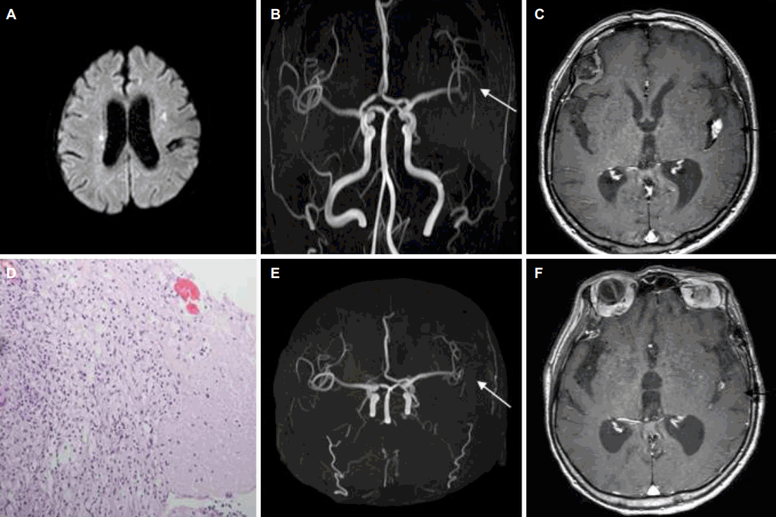
약 8개월 전부터 결핵수막염으로 항결핵제를 투약해온 77세 여자가 졸음상태가 지속되어 내원하였다. 확산강조영상에서 양측 대뇌반구의 다발뇌경색이 관찰되었고(Fig. A), 뇌자기공명영상에서 대뇌혈관의 혈관염을 시사하는 광범위한 조영제 증강과 좌측 중대뇌동맥 M2-분절의 협착이 보였다(Fig. B). 혈관벽-뇌자기공명영상을 시행하였으며, 실비우스수조에서 조영제 증강되는 결절이 중대뇌동맥을 둘러싸며 압박하였다(Fig. C). 결절의 현미경소견은 상피모양의 조직구와 림프구로 둘러싸인 중심괴사가 관찰되어 결핵육아종에 합당한 소견이었다(Fig. D). 기존 항결핵제와 병용하여 스테로이드 경구요법을 하였고, 9개월 후에 추적관찰한 혈관벽-뇌자기공명영상에서 좌측 중대뇌동맥 M2-분절협착이 호전되고 결절의 크기가 현저히 줄었다(Fig. E, F).
| J Korean Neurol Assoc > Volume 35(3); 2017 > Article |

|
Figure.
Brain images and biopsy findings of the patient. (A) Diffusion weighted image showed multiple cerebral infarct lesions in bilateral frontal white matter. (B) Brain CE MRA showed left MCA M2 stenosis (white arrow). (C) Vessel wall brain CE MRI revealed 1.2 cm-sized enhancing nodule at left sylvian cistern (black arrow). (D) The microscopic finding of enhancing nodule around left MCA revealed central necrosis surrounded by epithelioid histiocytes and lymphocytes (H&E stain, ×200). (E, F) Follow up vessel wall brain CE MRI showed slight improvement of luminal stenosis of left MCA M2 division (white arrow) and further decreased size of the enhancing mass-like lesion (black arrow). CE; contrast enhanced, MRA; magnetic resonance angiography, MCA; middle cerebral artery, MRI; magnetic resonance imaging, H&E; hematoxylin and eosin.
- TOOLS
 PDF Links
 PubReader
 ePub Link
 Full text via DOI
 Download Citation
 Print
Print


-
METRICS

-
- 0 Crossref
- 0 Scopus
- 5,115 View
- 132 Download
-

- Related articles
-
Traumatic Dissection of Middle Cerebral Artery with Intimal Flap2007 ;25(2)
- Editorial Office
-
(ZIP 03163) #1111, Daeil Bldg, 12, Insadong-gil, Jongno-gu, Seoul, Korea
Tel: +82-2-737-6530 Fax: +82-2-737-6531 E-mail: jkna@neuro.or.kr
Copyright © 2024 by Korean Neurological Association.

