

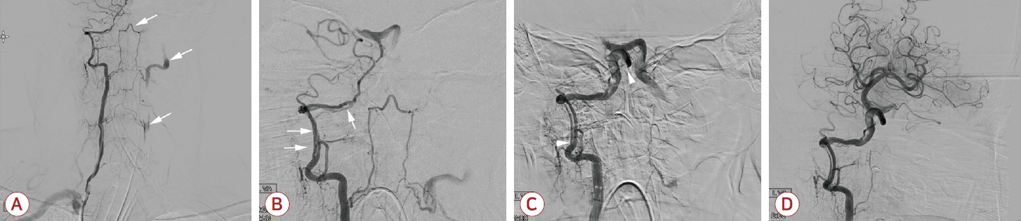
척추동맥박리는 급성 뇌바닥동맥폐색의 드문 원인으로 알려져 있다[1]. 박리된 내막에 의해 미만성 협착이 발생할 경우 특이도가 낮아[2] 혈관박리를 진단하기 어려울 뿐만 아니라 혈관 내 치료 시 폐색된 혈관으로 접근할 때 제한이 발생할 수 있다. 64세 남자가 2시간 전 갑자기 발생한 혼수와 경련으로 내원하여 시행한 컴퓨터단층촬영혈관조영술에서 뇌바닥동맥의 완전 폐색이 관찰되어 응급 혈전제거술을 시행하였다. 시술 중 헤파린첨가식염수(heparinized saline)의 지속적인 투여 외에 다른 항혈전제는 투약되지 않았다. 디지털감산혈관조영술에서 왼쪽 척추동맥은 기시부에서 완전 폐색되어 있으며, 오른쪽 척추동맥에서는 치상돌기 문합을 통해 상행혈류가 관찰되었다(Fig. A). 오른쪽 척추동맥조영술에서 척추동맥 V3-4 분절의 미만성 협착 때문에 폐색된 뇌바닥동맥까지 접근이 불가능하였다(Fig. B). 그러나 약 20분 간격을 두고 시행한 혈관조영술에서 처음에 관찰되지 않았던 오른쪽 척추동맥 V3-4 분절의 진성내강이 드러나며, 이중내강(double lumen), 내막절편(intimal flap)이 동반된 척추동맥박리가 관찰되었다(Fig. C). 이후 흡입 및 stentriever를 사용하여 혈전제거술을 시행하였고, 뇌기저동맥은 성공적으로 재개통되었으나, 오른쪽 V3-4 분절의 척추동맥박리는 계속 관찰되었다(Fig. D). 척추동맥박리에 대해서 추가적인 시술은 하지 않았다. 급성 뇌바닥동맥폐색에 대한 혈관 내 치료 시, 접근이 어려울 정도의 긴 분절성 미만성 협착은 척추동맥박리에 의한 진성내강 폐색을 고려해야 한다.
| J Korean Neurol Assoc > Volume 41(2); 2023 > Article |

|
REFERENCES
1. Mattle HP, Arnold M, Lindsberg PJ, Schonewille WJ, Schroth G. Basilar artery occlusion. Lancet Neurol 2011;10:1002-1014.


2. Gottesman RF, Sharma P, Robinson KA, Arnan M, Tsui M, Saber-Tehrani A, et al. Imaging characteristics of symptomatic vertebral artery dissection: a systematic review. Neurologist 2012;18:255-260.

Figure.
Digital subtraction angiography images of the patient. (A) Digital subtraction angiography images reveal occluded left vertebral artery (VA) supplied by anastomosis of the odontoid arch and muscular branches (white arrows). (B) Right vertebral angiogram shows complete occlusion of the basilar artery at the proximal 1/3 and long segmental stenosis of the V3-4 segment. Also, the appearance of a ‘double lumen’ was shown at the C2 to V4 segment, which was suspicious vertebral artery dissection (white arrows). (C) VA-gram demonstrates the dilated vascular structure with a double lumen and intimal flap which suggests of dissection on the right V3-4 segment. A dissected vertebral artery with a double lumen was demonstrated from just beyond the C2 curve to the vertebrobasilar junction (arrowheads). (D) Frontal angiogram shows full recanalization of the basilar artery after endovascular reperfusion therapy. However, the dissection of V3-4 segments has still remained.
- TOOLS
 PDF Links
 PubReader
 ePub Link
 Full text via DOI
 Download Citation
 Print
Print


-
METRICS

-
- 0 Crossref
- 0 Scopus
- 111 View
- 2 Download
-

- Related articles
- Editorial Office
-
(ZIP 03163) #1111, Daeil Bldg, 12, Insadong-gil, Jongno-gu, Seoul, Korea
Tel: +82-2-737-6530 Fax: +82-2-737-6531 E-mail: jkna@neuro.or.kr
Copyright © 2024 by Korean Neurological Association.

