

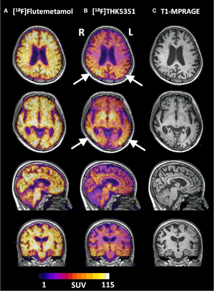
58세 여자가 약 3년 전 발생하여 서서히 진행하는 시각 증상과 시공간능력, 기억력을 포함한 인지기능저하로 내원하였다. 환자는 안과적 문제는 없었으나, 약 3년 전부터 바로 앞에 있는 물건도 잘 찾지 못하고, 걸어가다가 앞에 있는 물건을 보지 못하여 부딪히는 일이 자주 발생했으며, 서서 신발을 신는 일도 어려워졌다고 했다. 또한, 약 2년 전부터는 매일 다니는 등산길도 생소하게 생각하며, 익숙한 동네 마트에 갈 때도 매번 다른 층에 가거나 다른 입구로 들어가서 헤매는 일이 잦다고 하였다. 약 1-2년 전부터는 기억력이 현저히 떨어져 익숙한 기계를 작동하는 방법을 잊어버려서 전기밥솥, 세탁기, 핸드폰 사용 등이 어려워졌으며, 내원 당시에는 금방 들은 것도 잊어버리는 정도라고 하였다. 환자의 교육수준은 12년이었고 신경학적 진찰에서 동시실인증이 보였다. 신경심리검사에서 K-MMSE 는 14, CDR 1이었으며, Rey Complex Figure test에서 동일 연령, 학력의 정상인의 0.01 percentile 이하에 해당하는 등, SNSB II 검사의 거의 모든 항목에서 저하된 소견을 보였다. 상기 증상으로 환자는 후대뇌피질위축(posterior cortical atrophy)으로 진단받았으며, 새로운 tau PET tracer인 18F-THK5351 [1]와 amyloid PET tracer인 18F-Flutemetamol [2]를 이용한 PET 검사를 받았다. 18F-THK5351 PET 영상 결과, 전형적인 알츠하이머병 환자의 영상과 달리, 시각 증상을 주로 보인 임상 양상에 합당하게 양측(우측 > 좌측) 후두엽 및 측두엽, 두정엽을 포함한 일차시각 피질 및 연합시각피질에 타우단백질이 축적된 것을 확인할 수 있었으며, 양측 전두엽에도 경하게 침착된 소견이 보였다(Fig.). 18F-Flutemetamol PET 검사 결과, 전형적인 알츠하이머병의 amyloid PET 영상에서 보이는 바와 같이 연합피질에 걸쳐 전반적으로 아밀로이드단백질이 침착된 소견 외에도 특이적으로 후두엽에 아밀로이드단백질이 침착된 소견이 관찰되었다. 본 증례를 통하여 18F-THK5351을 이용한 tau PET 영상이 후대뇌피질위축 환자에서 임상 증상을 예민하게 반영한다는 것을 국내 최초로 보고한다.
| J Korean Neurol Assoc > Volume 34(4); 2016 > Article |

|
Acknowledgements
This study was supported by a grant of the Korea Health care Technology R&D Project through the Korean Health Industry Development Institute (KHIDI), funded by the Ministry of Health & Welfare, Republic of Korea (grant No. HI14C1135).
REFERENCES
1. Harada R, Okamura N, Furumoto S, Furukawa K, Ishiki A, Tomita N, et al. 18F-THK5351: A Novel PET Radiotracer for imaging neurofibrillary pathology in alzheimer disease. J Nucl Med 2016;57:208-214.


2. Mountz JM, Laymon CM, Cohen AD, Zhang Z, Price JC, Boudhar S, et al. Comparison of qualitative and quantitative imaging characteristics of [11C]PiB and [18F]flutemetamol in normal control and Alzheimer's subjects. Neuroimage Clin 2015;9:592-598.

Figure.
(A) Axial, sagittal, and coronal images of 18F-Flutemetamol PET, (B) 18F-THK5351 PET, and (C) MR (T1-MPRAGE) images of the patient. (A) Flutmetamol retention was shown in the bilateral occipital cortex as well as frontal, parietal, lateral temporal cortices and precuneus. (B) Significant THK retention was found in the bilateral parietal, temporo-occipital areas (Rt > Lt) (arrows) and mild THK retention was shown in bilateral frontal areas. (C) T1 MPRAGE images showed diffuse brain atrophy, especially severe finding in temporo-occipital area (Rt > Lt). Color bar shows SUV ranges. MPRAGE; magnetization prepared rapid acquisition gradient echo, SUV; standard uptake value, Rt; right, Lt; left.
- TOOLS
 PDF Links
 PubReader
 ePub Link
 Full text via DOI
 Download Citation
 Print
Print


-
METRICS

-
- 0 Crossref
- 0 Scopus
- 7,519 View
- 219 Download
- Related articles
- Editorial Office
-
(ZIP 03163) #1111, Daeil Bldg, 12, Insadong-gil, Jongno-gu, Seoul, Korea
Tel: +82-2-737-6530 Fax: +82-2-737-6531 E-mail: jkna@neuro.or.kr
Copyright © 2026 by Korean Neurological Association.

