Roles of Diagnostic Cerebral Angiography and High-resolution Vessel-wall Imaging in Evaluating Basilar Artery Perforators: A Case of Bilateral Midbrain Infarction
- Hokyu Kim, MD, Jung Hoon Han, MD, Chi Kyung Kim, MD, PhD, Kyungmi Oh, MD, PhD, Keon-Joo Lee, MD*, Sang-Il Suh, MD, PhDa,*
нҳҲкҙҖмЎ°мҳҒмҲ л°Ҹ кі н•ҙмғҒлҸ„ лҮҢнҳҲкҙҖлІҪ MRIлҘј мқҙмҡ©н•ҳм—¬ 진лӢЁлҗң лӢЁмқј кҙҖнҶөлҸҷл§ҘмңјлЎң л°ңмғқн•ң м–‘мёЎ л°©м •мӨ‘ мӨ‘лҮҢкІҪмғү
- к№Җнҳёк·ң, н•ңм •нӣҲ, к№Җм№ҳкІҪ, мҳӨкІҪлҜё, мқҙкұҙмЈј*, м„ңмғҒмқјa,*
- Received November 6, 2024; В В В Revised January 22, 2025; В В В Accepted February 3, 2025;
- ABSTRACT
-
Bilateral midbrain infarctions are often associated with basilar artery (BA) steno-occlusion, but identifying the stroke etiology is difficult when large vessels appear normal. We present a 90-year-old female with wall-eyed bilateral internuclear ophthalmoplegia. Initial diffusion-weighted imaging showed subtle bilateral midbrain lesions, while computed tomography angiography produced normal findings. Vessel-wall imaging and diagnostic angiography identified an abnormal single perforator from the distal BA supplying both sides. This case highlights the importance of these techniques in detecting perforator abnormalities in stroke with unclear etiology.
Ischemic stroke involving the bilateral brainstem needs to be differentiated from metabolic brain disorders, toxic brain disorders, and central nervous system tumors [1]. Bilateral midbrain infarctions are commonly associated with steno-occlusion of the basilar artery (BA) and bilateral vertebral arteries (VAs), along with anatomical variants of the BA as contributing causes [2]. Identifying the etiology of stroke through conventional imaging techniques such as magnetic resonance angiography and computed tomography angiography (CTA) can be challenging when large vessels appear normal and the condition arises from deep perforator problems. We report a rare case of pure bilateral midbrain infarction without large-vessel abnormalities, in which the pathology of a single deep perforator was detected by transfemoral cerebral angiography (TFCA) and high-resolution vessel-wall imaging (VWI).
- CASE
- CASE
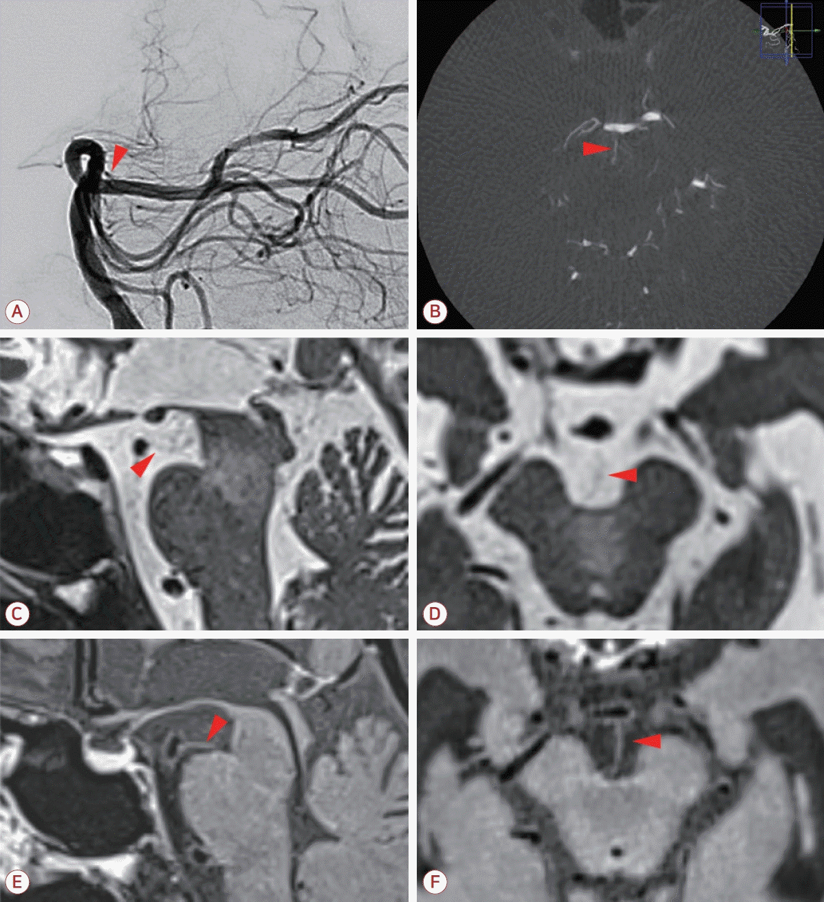
A 90-year-old female with hypertension was admitted to the emergency room with acute-onset dysarthria and dizziness persisting for 6 hours without any other neurological deficits. Initial diffusion-weighted imaging (DWI) revealed subtle diffusion-restricted lesions in the bilateral anteromedial portions of the midbrain tegmentum (Fig. 1-A, B). Computed tomography perfusion revealed cerebral hypoperfusion extending ventral to the aqueduct in the bilateral paramedian part of the midbrain (Fig. 1-C). CTA revealed a fetal variant of the right posterior cerebral artery (PCA) without significant stenosis in other intracranial arteries (Fig. 1-D, E). Informed consent was obtained from the patient for publishing this case report, and the local Institutional Review Board (IRB) approved its design (IRB No. 2024GR0042).Two days after admission, the patientвҖҷs neurological status deteriorated, as evidenced by more-prominent bilateral paramedian lesions in follow-up DWI (Fig. 1-F). This deterioration was associated with clinical symptoms of wall-eyed bilateral internuclear ophthalmoplegia and ataxia affecting both upper and lower extremities (Fig. 1-G). TFCA was used to further assess the lesion attributes and hidden vascular pathologies. A deep perforator of the distal BA was identified in a conventional angiography image (Fig. 2-A). Multiplanar reformatting imaging revealed a single common perforator entering the interpeduncular fossa, which appeared to be associated with the ischemic lesion (Fig. 2-B). VWI performed to obtain additional information about the vascular abnormalities confirmed the detailed trajectory of the perforator from the distal BA directly toward the ischemic lesion (Fig. 2-C, D) and revealed abnormal vessel-wall enhancement of this perforator (Fig. 2-E, F).Serum laboratory findings were normal, including the levels of vitamins B1 and B12, blood urea nitrogen, ammonia, glucose, and electrolytes, which ruled out any other causes of the bilateral midbrain lesions. Furthermore, no other potential sources of stroke were found, leading to the final diagnosis of bilateral pure midbrain infarction resulting from an abnormality in the single perforator of the distal BA. The neurological state of the patient gradually improved over 2 weeks, and she was discharged in a stable condition.
- DISCUSSION
- DISCUSSION
This was a rare case of bilateral pure midbrain infarction caused by an abnormality in the single common perforator of the distal BA supplying both sides of the paramedian part of the midbrain. Bilateral midbrain infarction can result from severe stenosis or occlusion of the BA and VAs, but the occurrence of pure midbrain infarction without the involvement of other structures such as the thalamus or pontine is uncommon [3,4]. The blood supply to the paramedian and central parts of the midbrain is typically associated with perforators from the PCA, such as the thalamoperforating, peduncular, and mesencephalic arteries [5]. The well-known rostral group of perforators from the distal BA typically extend into the interpeduncular fossa, but their direct involvement in supplying the paramedian part of the midbrain, as seen in the present case, has been rarely documented [5,6]. Therefore, pure bilateral midbrain infarction caused by a single deep perforator abnormality without significant large-vessel involvement can be difficult to diagnose. TFCA and high-resolution VWI offered important insights into the underlying pathology of the stroke in the present case.Diagnostic cerebral angiography is well known for providing detailed information about intracranial arteries, and VWI is widely recognized for its accurate assessments of the wall pathology and pathways of large arteries [7]. The present case demonstrates how the combined use of diagnostic cerebral angiography and high-resolution VWI also enables 1) the identification of specific deep perforators contributing to the lesion from among numerous deep perforators, 2) the precise mapping of their trajectories to visualize the correlation with the affected parenchymal lesions, and 3) the detection of their abnormal vessel-wall enhancement. This case thus provides additional diagnostic insights for determining the etiology of stroke caused by unusual deep perforator problems, which may not be fully explained by simple conventional techniques.
FigureВ 1.
Simple imaging and ophthalmologic findings of the patient (A, B) Initial diffusion-weighted imaging (DWI) showed subtle diffusion restriction in the bilateral anteromedial portions of the midbrain. (C) A delayed time-to-maximum (Tmax) was observed in computed tomography perfusion that extended ventral to the aqueduct of the bilateral midbrain. (D, E) Computed tomography angiography did not reveal any significant stenosis in the posterior circulation, including the basilar artery and the posterior cerebral artery. (F) Follow-up DWI showed acute ischemic lesions in the central portion of the midbrain, ventral to the aqueduct and affecting the paramedian structures bilaterally; these lesions were more prominent than in the initial imaging. (G) Eye movements demonstrated primary gaze exotropia, bilateral adduction impairment on the lateral gaze (red arrows), vertical gaze impairment, and bilateral abduction nystagmus, confirming the presence of wall-eyed bilateral internuclear ophthalmoplegia.

FigureВ 2.
Diagnostic cerebral angiography and vessel-wall imaging of the patient (A) Lateral view of the left vertebral artery injection showing a rostral perforator of the basilar artery (red arrowhead). (B) Multiplanar reformatting image showing a specific single common perforator of the distal BA entering the interpeduncular fossa (red arrowhead). (C, D) Axial T2-weighted sampling perfection with application optimized contrast using different flip-angle evolution (SPACE) sequence showing the trajectory of the perforator from the distal BA toward the long-extending ischemic lesion (red arrowhead). (E, F) Postcontrast T1-weighted delay alternating with nutation for tailored excitation (DANTE)-SPACE image showing abnormal wall enhancement of the perforator (red arrowhead).

- REFERENCES
- REFERENCES
- 1. de Oliveira AM, Paulino MV, Vieira AP, McKinney AM, da Rocha AJ, Dos Santos GT, et al. Imaging patterns of toxic and metabolic brain disorders. Radiographics 2019;39:1672-1695.
[Article] [PubMed]2. Spengos K, Wohrle JC, Tsivgoulis G, Stouraitis G, Vemmos K, Zis V. Bilateral paramedian midbrain infarct: an uncommon variant of the вҖңtop of the basilarвҖқ syndrome. J Neurol Neurosurg Psychiatry 2005;76:742-743.
[Article] [PubMed] [PMC]3. Kim JS, Kim J. Pure midbrain infarction: clinical, radiologic, and pathophysiologic findings. Neurology 2005;64:1227-1232.
[Article] [PubMed]4. Lazzaro NA, Wright B, Castillo M, Fischbein NJ, Glastonbury CM, Hildenbrand PG, et al. Artery of percheron infarction: imaging patterns and clinical spectrum. AJNR Am J Neuroradiol 2010;31:1283-1289.
[Article] [PubMed] [PMC]5. Vogels V, Dammers R, van Bilsen M, Volovici V. Deep cerebral perforators: anatomical distribution and clinical symptoms: an overview. Stroke 2021;52:e660-e674.
[PubMed]
