Mobile Thrombus within Carotid Artery Stent
- Hyun Goo Kang, MD, In Sung Choo, MD, Bum Joon Kim, MDa, Jin Sung Cheong, MDb
кІҪлҸҷл§Ҙ мҠӨн…җнҠё лӮҙл¶Җм—җм„ң ліҙмқё мӣҖм§ҒмқҙлҠ” нҳҲм „
- к°•нҳ„кө¬, 추мқём„ұ, к№ҖлІ”мӨҖa, м •м§„м„ұb
- Received February 5, 2016; В В В Revised March 23, 2016; В В В Accepted March 23, 2016;
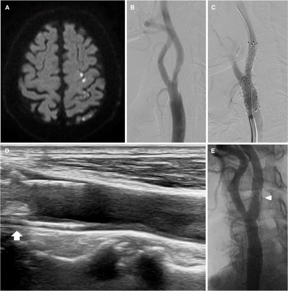
кі нҳҲм••кіј лӢ№лҮЁлі‘мқҙ мһҲлҚҳ 73м„ё лӮЁмһҗк°Җ 1л…„ м „ мўҢмёЎ мӨ‘лҢҖлҮҢлҸҷл§ҘмҳҒм—ӯм—җ кёүм„ұ лҮҢкІҪмғүкіј(Fig. A) мўҢмёЎ кІҪлҸҷл§Ҙ к·јмң„л¶Җм—җ нҳ‘м°©(52.5%)кіј кІҪкі„к°Җ л¶Ҳк·ңм№ҷн•ң нҳҲм „мңјлЎң(Fig. B) кІҪлҸҷл§ҘмҠӨн…җнҠёлҘј мӮҪмһ…н•ҳмҳҖлӢӨ(Fig. C). 1л…„ нӣ„ мӢңн–үн•ң кІҪлҸҷл§ҘмҙҲмқҢнҢҢм—җм„ң мҠӨн…җнҠё лӮҙл¶Җм—җ мһ¬нҳ‘м°©кіј мң лҸҷм„ұ нҳҲм „(mobile thrombus)мқҙ ліҙмҳҖкі (Fig. D) лҮҢнҳҲкҙҖмЎ°мҳҒмҲ м—җм„ңлҠ” мҠӨн…җнҠёлӮҙмһ¬нҳ‘м°©мқҙ мһҲм—ҲлӢӨ(Fig. E). лҮҢнҳҲкҙҖмЎ°мҳҒмҲ м—җм„ң ліҙмқҙлҠ” мҠӨн…җнҠёлӮҙмһ¬нҳ‘м°©мқ„ кІҪлҸҷл§ҘмҙҲмқҢнҢҢлЎң ліҙм•ҳмқ„ л•Ң нҳҲм „мқҳ нҳ•нғңлҘј 분лӘ…н•ҳкІҢ нҷ•мқён• мҲҳ мһҲм—ҲлӢӨ. мҠӨн…җнҠё мӮҪмһ… нӣ„ мҠӨн…җнҠёлӮҙмһ¬нҳ‘м°©мқ„ 진лӢЁн•ҳкё° мң„н•ҙ лҮҢнҳҲкҙҖмЎ°мҳҒмҲ , CTнҳҲкҙҖмЎ°мҳҒмҲ , кІҪлҸҷл§ҘмҙҲмқҢнҢҢлҘј мӮ¬мҡ©н•ңлӢӨ[1]. нҠ№нһҲ кІҪлҸҷл§ҘмҙҲмқҢнҢҢлҠ” 비침мҠөм Ғмқё кІҖмӮ¬лЎң к°„нҺён•ҳкі м•Ҳм „н•ҳкІҢ л°ҳліөм ҒмңјлЎң кІҖмӮ¬лҘј н• мҲҳ мһҲм–ҙ[2], мҠӨн…җнҠё нӣ„ мғүм „ мң„н—ҳмқҙ лҶ’мқҖ нҳҲм „мқҳ нҠ№м„ұмқ„ нҷ•мқён• мҲҳ мһҲкі , м№ҳлЈҢл°©н–Ҙ кІ°м •м—җлҸ„ лҸ„мӣҖмқҙ лҗңлӢӨ.
Figure.
(A) Acute multiple scattered infarction in left middle cerebral artery territory on diffusion weighted image. (B) Left proximal carotid artery showing irregular marginal thrombosis and stenosis (52.5%) at transfemoral cerebral angiography (TFCA). (C) Immediately after deployment of carotid artery stent. (D) In-stent thrombosis at follow-up carotid ultrasonography, one year after stenting (arrow) (E) In-stent restenosis at follow up TFCA, one year after stenting (arrow head).