항암 치료 유발 혈소판감소증 환자에서 두통과 안구출혈로 발현한 중뇌 주위 지주막하출혈
Perimesencephalic Subarachnoid Hemorrhage Presenting with Headache and Ocular Hemorrhage in a Patient with Chemotherapy-induced Severe Thrombocytopenia
Article information
Trans Abstract
A 26-year-old female with severe thrombocytopenia (platelet count <20,000/μL) following combination chemotherapy (cisplatin, paclitaxel, pembrolizumab, and bevacizumab) for cervical cancer presented with new-onset headache and ocular hemorrhage. Perimesencephalic subarachnoid hemorrhage (pmSAH) was identified, which was subsequently complicated by disproportionate cerebral vasospasm and transient oculomotor nerve palsy. This case demonstrates that pmSAH, which is generally considered a benign venous hemorrhage, can follow an unusual course in the setting of profound bleeding diathesis and chemotherapy-induced vascular injury.
Perimesencephalic subarachnoid hemorrhage (pmSAH) is classically considered a benign form of nonaneurysmal subarachnoid hemorrhage (SAH) that is typically characterized by a mild clinical presentation and a very low risk of delayed complications such as cerebral vasospasm [1,2]. However, the presence of severe underlying systemic conditions can markedly alter the clinical course of pmSAH. Severe thrombocytopenia (platelet count <20,000/μL), particularly following chemotherapy, not only increases the risk of intracranial hemorrhage but may also alter its presentation and progression [3]. Furthermore, systemic exposure to multiagent chemotherapy, especially vascular endothelial growth factor (VEGF) inhibitors such as bevacizumab, can compromise the integrity of the vascular endothelium and result in significant vascular fragility [4,5].
We describe a 26-year-old female with chemotherapy-induced severe thrombocytopenia who developed pmSAH with a complicated atypical course. Following the onset of headache and sentinel ocular hemorrhage she developed disproportionate cerebral vasospasm and transient oculomotor nerve palsy. This case highlights how the combination of profound bleeding diathesis and chemotherapy-induced endothelial dysfunction can transform a generally benign venous hemorrhage into an unusual and potentially aggressive clinical entity.
CASE REPORT
A 26-year-old female with advanced cervical adenocarcinoma was referred to our neurology department for evaluation of a newly developed headache. She had been hospitalized for the conservative management of severe pancytopenia that had developed following a recent history of chemotherapy. She had received combination chemotherapy including cisplatin, paclitaxel, pembrolizumab, and bevacizumab 2 months prior to the onset of her headache, and 1 month prior to symptom onset she had been maintained on cisplatin monotherapy.
On the day of symptom onset she acutely developed a persistent headache and posterior neck pain with a pain intensity of approximately 5 on a numeric rating scale from 0 to 10. She had no prior history of migraine or tension-type headache. The pain was consistently exacerbated by neck movements, including forward flexion and extension, suggesting meningeal irritation. The headache was accompanied by right-sided conjunctival hemorrhage and intermittent fever spikes up to approximately 38℃ that occurred once or twice daily, but without nausea, vomiting, dizziness, or other neurological symptoms. Other vital signs remained stable.
A neurological examination revealed an alert and cooperative mental status with intact cranial nerve function, symmetric motor and sensory findings, and preserved cerebellar function. A laboratory evaluation at presentation revealed severe pancytopenia, with extreme thrombocytopenia (platelet count <5,000/μL). Serial blood cultures were positive for Gram-positive organisms, and she received intravenous cefepime and vancomycin as treatment for neutropenic fever. An ophthalmological examination revealed right-sided subconjunctival hemorrhage, Roth spots, and bilateral retinal hemorrhages (Fig. 1).
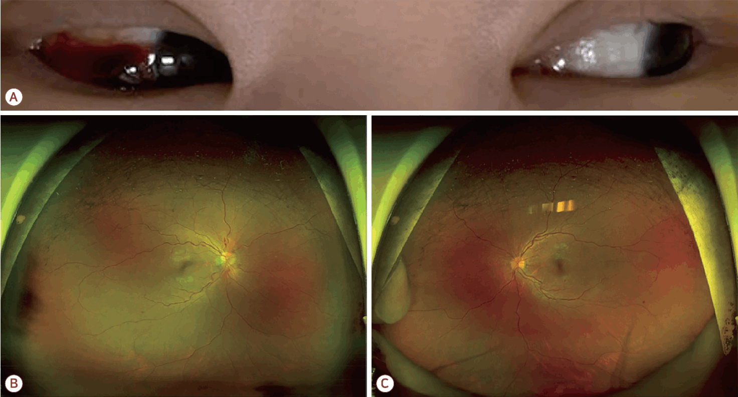
Findings for ocular hemorrhages. (A) Clinical photograph demonstrating right-sided subconjunctival hemorrhage. (B) Fundus photograph of the right eye showing multiple retinal hemorrhages and Roth spots. (C) Fundus photograph of the left eye demonstrating multiple retinal hemorrhages.
Given the combination of severe thrombocytopenia and ocular hemorrhage, intracranial hemorrhage was considered the primary diagnostic concern, with secondary consideration of central nervous system infection. Brain magnetic resonance imaging with contrast enhancement demonstrated small areas of SAH involving the interpeduncular, prepontine, and suprasellar cisterns as well as the anterior interhemispheric fissure and left frontal sulci (Fig. 2). Multifocal pachymeningeal enhancement was observed along the bilateral temporal and parietal convexities, suggesting inflammatory changes, while meningeal metastasis was considered less likely. Subsequent computed tomography angiography revealed no intracranial aneurysm, but demonstrated diffuse narrowing of both M1 segments of the middle cerebral artery, raising concern about cerebral vasospasm (Fig. 3). A lumbar puncture and digital subtraction angiography (DSA) were deferred due to the severe thrombocytopenia and high bleeding risk. A neurosurgical consultation favored a diagnosis of nonaneurysmal pmSAH. Conservative management was initiated with intravenous mannitol (80 mL every 8 hours) and intravenous nimodipine (50 mg/day).
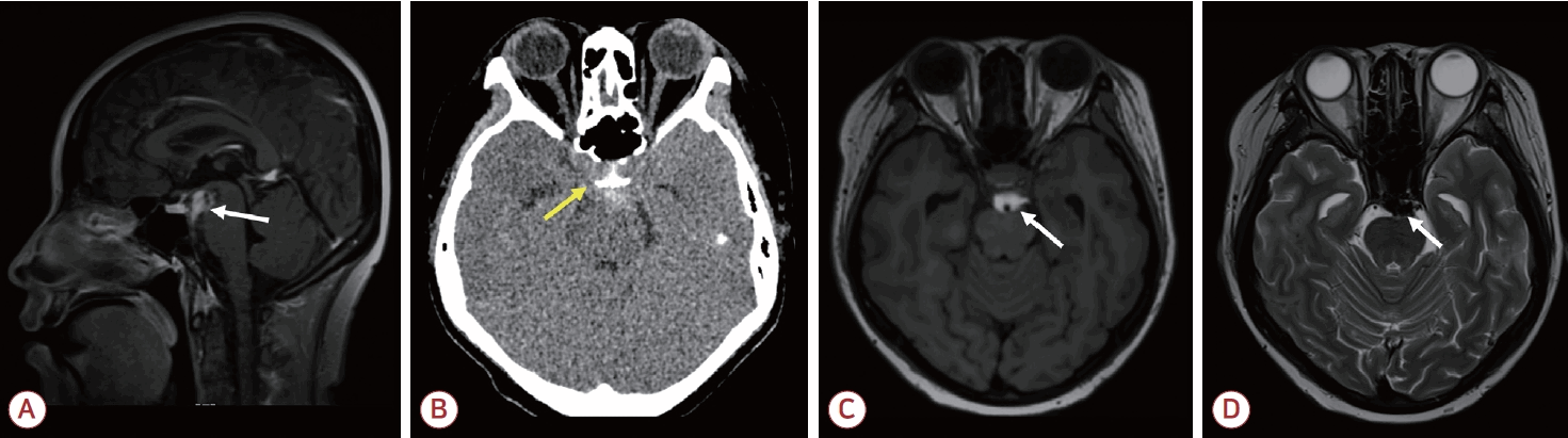
Brain imaging findings for pmSAH. (A) Sagittal T1-weighted magnetic resonance imaging (MRI) revealed hyperintense signals within the interpeduncular and prepontine cisterns (white arrow). (B) Axial noncontrast computed tomography (CT) revealed hyperdense signals in the perimesencephalic cisterns (yellow arrow). (C) Axial T1-weighted MRI showed the corresponding subarachnoid hemorrhage with a high signal intensity (white arrow). (D) Axial T2-weighted MRI demonstrated low signal intensities within the perimesencephalic cisterns, consistent with hemorrhage (white arrow). pmSAH, perimesencephalic subarachnoid hemorrhage.
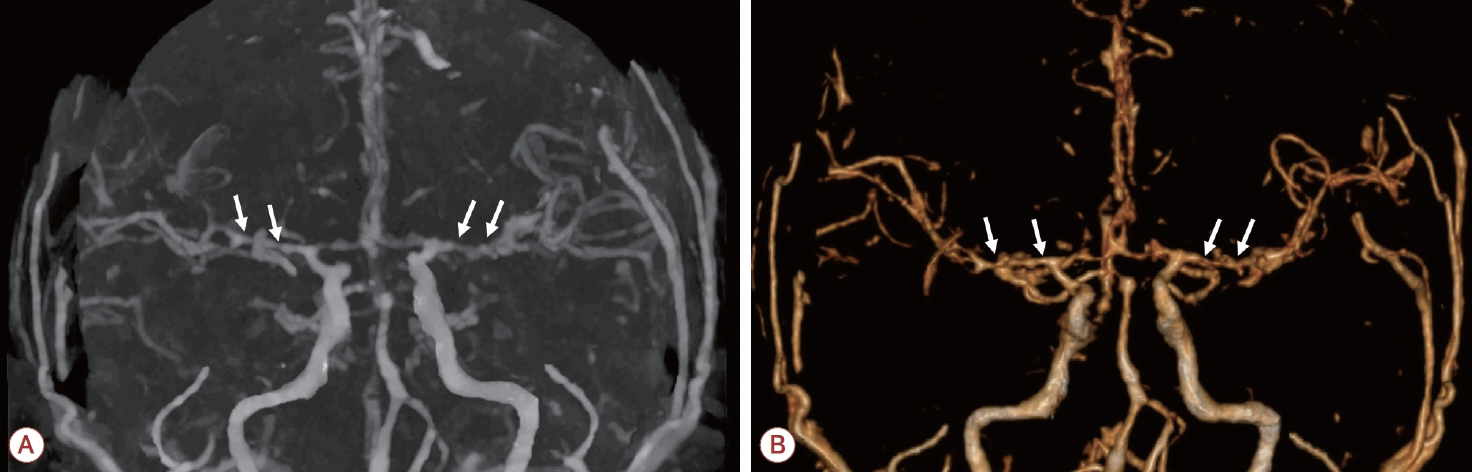
Findings from computed tomography (CT) angiography. (A) Axial CT angiography demonstrated diffuse narrowing of both M1 segments of the middle cerebral arteries (white arrows). (B) Three-dimensional reconstructed angiography image confirms bilateral M1 segment narrowing (white arrows) without evidence of intracranial aneurysm.
The patient experienced worsening nausea and vomiting during this conservative management, and the headache pattern evolved from occipital pain to bilateral temporal headache. At 7 days after the onset of the headache the patient developed transient left ptosis and binocular diplopia suggestive of left oculomotor nerve palsy, without accompanying motor or sensory deficits. This newly developed neurological deficit was evaluated by performing follow-up brain magnetic resonance angiography (MRA), which demonstrated no increase in the hemorrhage volume and no worsening of the cerebral vasospasm, with no additional lesions identified. Reassured by these stable radiological findings, we maintained the ongoing conservative treatment with transfusion support and intravenous mannitol and nimodipine.
At 10 days after the initial headache (i.e., 3 days after the onset of the cranial nerve palsy) her ptosis, diplopia, and headache began to show significant clinical improvements. She became completely pain-free without analgesics at 2 weeks after symptom onset. Furthermore, follow-up brain MRA performed 12 days after the headache onset confirmed the gradual resolution of the pmSAH and that the multifocal vasospasm was improving. Intravenous nimodipine was maintained for 14 days and then transitioned to oral nimodipine (60 mg five times daily), which was continued for an additional week. The patient was subsequently transferred to another tertiary hospital for ongoing oncological management.
DISCUSSION
This case illustrates several important clinical implications of nonaneurysmal pmSAH in the setting of chemotherapy-induced severe thrombocytopenia. While pmSAH is generally considered a venous hemorrhage with a favorable prognosis [6], our patient developed an atypical course involving concurrent ocular hemorrhage, disproportionate cerebral vasospasm, and transient oculomotor nerve palsy [2,7].
The ocular hemorrhage in this patient warrants careful interpretation. Terson syndrome is classically characterized by intraocular hemorrhage occurring in association with abrupt elevation of the intracranial pressure [8]. While our patient presented with ocular hemorrhage concurrently with SAH, pmSAH typically does not cause dramatic pressure increases. Therefore, the extensive ocular bleeding observed in this case likely represents a combined manifestation, triggered by the SAH but markedly exacerbated by the underlying profound bleeding diathesis and vascular fragility, rather than being a purely pressure-driven phenomenon.
Regarding the vascular complications, we attribute the unusually pronounced cerebral vasospasm to a multifactorial vascular insult. Beyond the mechanical effect of the subarachnoid blood, the patient’s recent history of multiagent chemotherapy, including the VEGF inhibitor such as bevacizumab, likely compromised the integrity of the vascular endothelium [4,5,9]. This endothelial dysfunction coupled with profound refractory thrombocytopenia predisposed the cerebral vessels to enhanced vasospasm.
Furthermore, the mechanism underlying oculomotor nerve palsy in pmSAH is distinct from the direct pulsatile arterial compression that is typically seen in aneurysmal SAH. We suspect that while our patient exhibited a multifactorial transient palsy, it primarily resulted from the direct localized mass effect of the dense hematoma accumulating in the interpeduncular and perimesencephalic cisterns where the oculomotor nerve exits the brainstem [7]. Secondary mechanisms including local inflammatory and toxic effects from subarachnoid blood breakdown products, as well as localized microvascular ischemia or vasospasm of the vasa nervorum, likely also contributed to the observed neural dysfunction [7].
This study had limitations. The severe pancytopenia and high risk of bleeding precluded invasive diagnostic modalities such as DSA, lumbar puncture, and routine transcranial Doppler monitoring. Additionally, while the acute neurovascular complications stabilized in our patient, she died from sepsis 3 months later due to her severely immunocompromised state.
In conclusion, the combination of severe thrombocytopenia and chemotherapy-induced vascular injury can critically alter the natural course of intracranial hemorrhages. This case demonstrates that profound vascular fragility and endothelial dysfunction, potentiated by VEGF inhibitors, can result in pmSAH deviating from its typically benign course to present with disproportionate complications such as vasospasm and cranial nerve palsy. Clinicians should anticipate such atypical courses and maintain vigilant neurovascular monitoring in highly vulnerable oncological patients.