가슴샘종 절제술 11년 후 발생한 굿(Good)증후군 환자의 진행다초점백질뇌병증
Progressive Multifocal Leukoencephalopathy in a Patient with Good Syndrome 11 Years after Thymectomy
Article information
Trans Abstract
Progressive multifocal leukoencephalopathy (PML) occurs in immunocompromised patients. We report a 60-year-old woman with rapidly progressive aphasia and cognitive decline 11 years after thymectomy. Brain magnetic resonance imaging revealed asymmetric, non-enhancing confluent white matter lesions. Laboratory tests showed hypogammaglobulinemia, reduced B cells, and a reversed CD4/CD8 ratio, consistent with Good syndrome. John Cunningham virus DNA was detected in cerebrospinal fluid, confirming PML. This case highlights that PML can occur in patients with Good syndrome even years after remote thymectomy.
진행다초점백질뇌병증(progressive multifocal leukoencephalopathy, PML)은 존커닝햄바이러스(John Cunningham virus, JCV)의 활성화로 인해 중추신경계의 희소돌기아교세포가 감염되어 발생하는 치명적인 탈수초질환이다[1,2]. 주로 장기 이식 후 면역억제제 사용, 인체면역결핍바이러스(human immunodeficiency virus, HIV) 감염, 림프종과 같은 심각한 면역 결핍 상태에서 발생하며, 일반적으로 무증상으로 잠복 감염 상태로 존재하지만 세포면역기능이 저하된 상황에서 재활성화되어 중추신경계를 침범할 수 있다.
가슴샘은 T림프구의 분화와 성숙이 이루어지는 1차 림프기관으로 중추면역관용의 형성과 면역계의 정상적인 조절에 중요한 역할을 한다. 가슴샘종(thymoma)이 발생하면 이러한 면역 조절 과정에 이상이 생길 수 있으며, 일부 환자에서는 체액 및 세포면역기능 저하가 동반될 수 있다. 특히 저감마글로불린혈증과 B세포 감소, T세포의 기능 저하를 특징으로 하는 드문 면역 결핍 질환인 굿증후군(Good syndrome)이 발생할 수 있으며, 이로 인하여 다양한 기회 감염이 나타날 수 있다[3].
저자들은 과거 가슴샘종 절제술을 받은 환자에서 수술 후 11년이 경과한 시점에 PML이 진단된 증례를 경험하였기에 문헌 고찰과 함께 보고하고자 한다.
증 례
60세 우즈베키스탄 국적의 여자가 점차 진행하는 조음장애, 언어장애 및 인지기능 저하로 병원에 왔다. 약 1개월 전부터 착어증(paraphasia)이 발생하였고 점차 악화되어 진찰 당시에는 운동실어증과 함께 알아듣기 힘들 정도의 심한 조음장애를 보였으며 전신 쇠약도 같이 진행된 상태였다. 의욕 저하로 인한 식사량 감소로 최근 1개월간 약 10 kg의 체중 감소가 동반되었고 신경계 진찰에서 사지 근력은 Medical Research Council 등급 4로 저하되어 있었다. 과거력으로 갑상샘기증저하증으로 레보티록신을 복용 중이었다. 16년 전 건강 검진에서 세로칸 덩이가 발견되어 경과 관찰하던 중 11년 전 가슴샘종 절제술을 받았다(Fig. 1-A). 당시 조직병리 결과는 침습가슴샘종으로 세계보건기구(World Health Organization, WHO) type AB, Masaoka stage IIA였으며 수술 후 추가적인 항암 치료나 방사선 치료 없이 정기적으로 추적 관찰 중이었다.
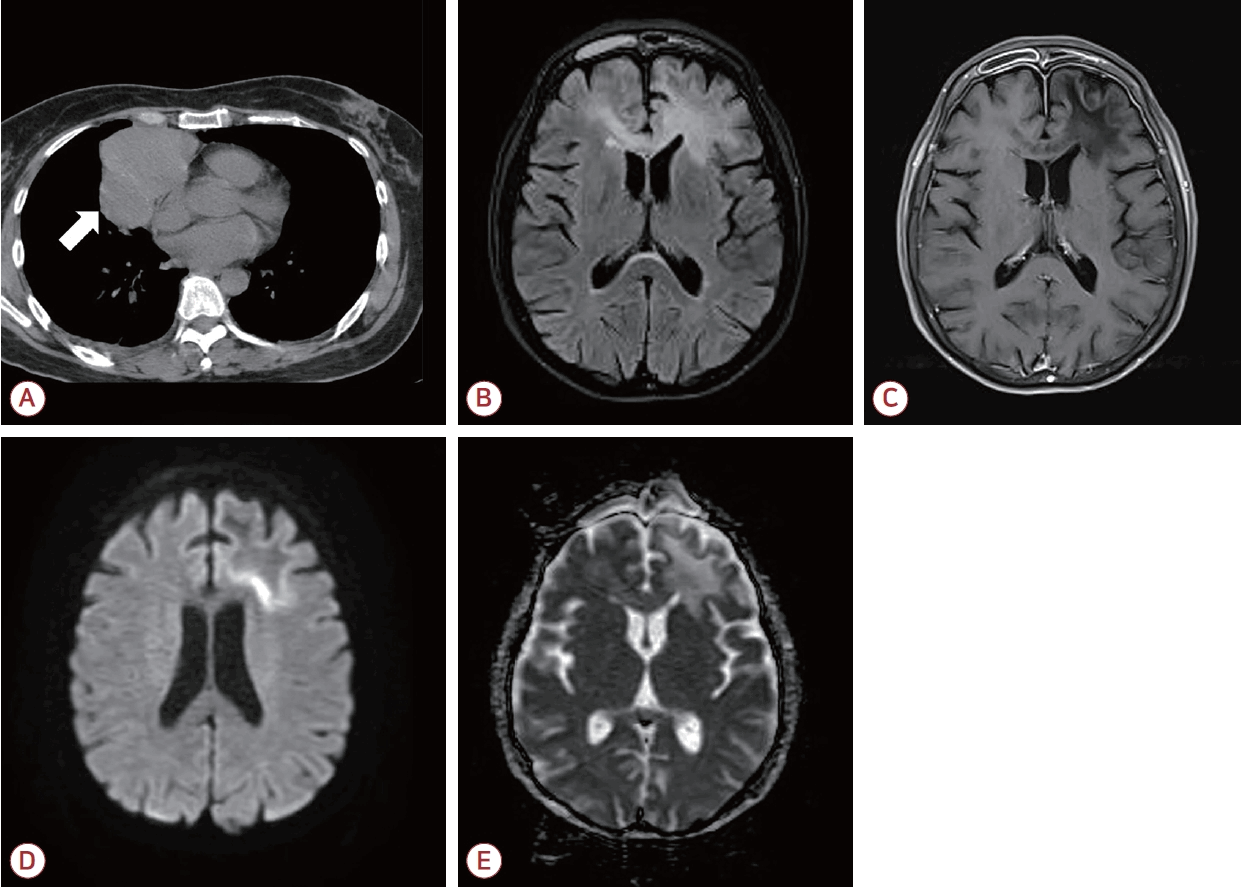
Chest computed tomography and brain magnetic resonance imaging findings. (A) Chest computed tomography performed 11 years earlier reveals an anterior mediastinal mass (white arrow), consistent with a thymoma. (B) Axial fluid-attenuated inversion recovery image of the brain obtained 10 days after symptom onset demonstrates asymmetric, confluent hyperintense lesions involving the subcortical and deep white matter of the bilateral frontal lobes and the genu of the corpus callosum. (C) Gadolinium-enhanced T1-weighted image shows no significant enhancement within the lesions. (D, E) Diffusion-weighted imaging (D) and the apparent diffusion coefficient map (E) demonstrate partial diffusion restriction within the lesions.
증상 발생 10일 후 시행한 뇌자기공명영상(magnetic resonance imaging, MRI)에서 피질(cortex)은 보존되었으나 양측 전두엽에서 비대칭으로 피질하백질, 심부백질, 뇌들보무릎(genu of corpus callous), 속섬유막앞다리(anterior limb of internal capsule) 및 좌측 측두엽 피질하백질에 T2강조영상과 액체감쇠역전회복(fluid-attenuated inversion recovery, FLAIR) 영상에서 연속적인 고신호강도 병터가 관찰되었고 T1강조영상에서는 저신호강도를 보였다(Fig. 1-B-E). 일부 병터에서는 확산 제한(diffusion restriction)이 보였으나 조영증강 소견은 없었다. 추적 관찰한 뇌MRI에서는 기존 병터의 범위가 확장되었으며 양측 후두엽 백질에서 T2강조영상 및 FLAIR에서 고신호강도 병터가 새롭게 관찰되었다(Fig. 2-A-C). 자기공명분광(magnetic resonance spectroscopy)에서는 N-아세틸아스파트산(N-acetylaspartate) 최고점 감소, 콜린 최고점의 경미한 증가 및 지질 최고점의 증가가 관찰되어 탈수초를 시사하는 소견이 보였다(Fig. 2-D).
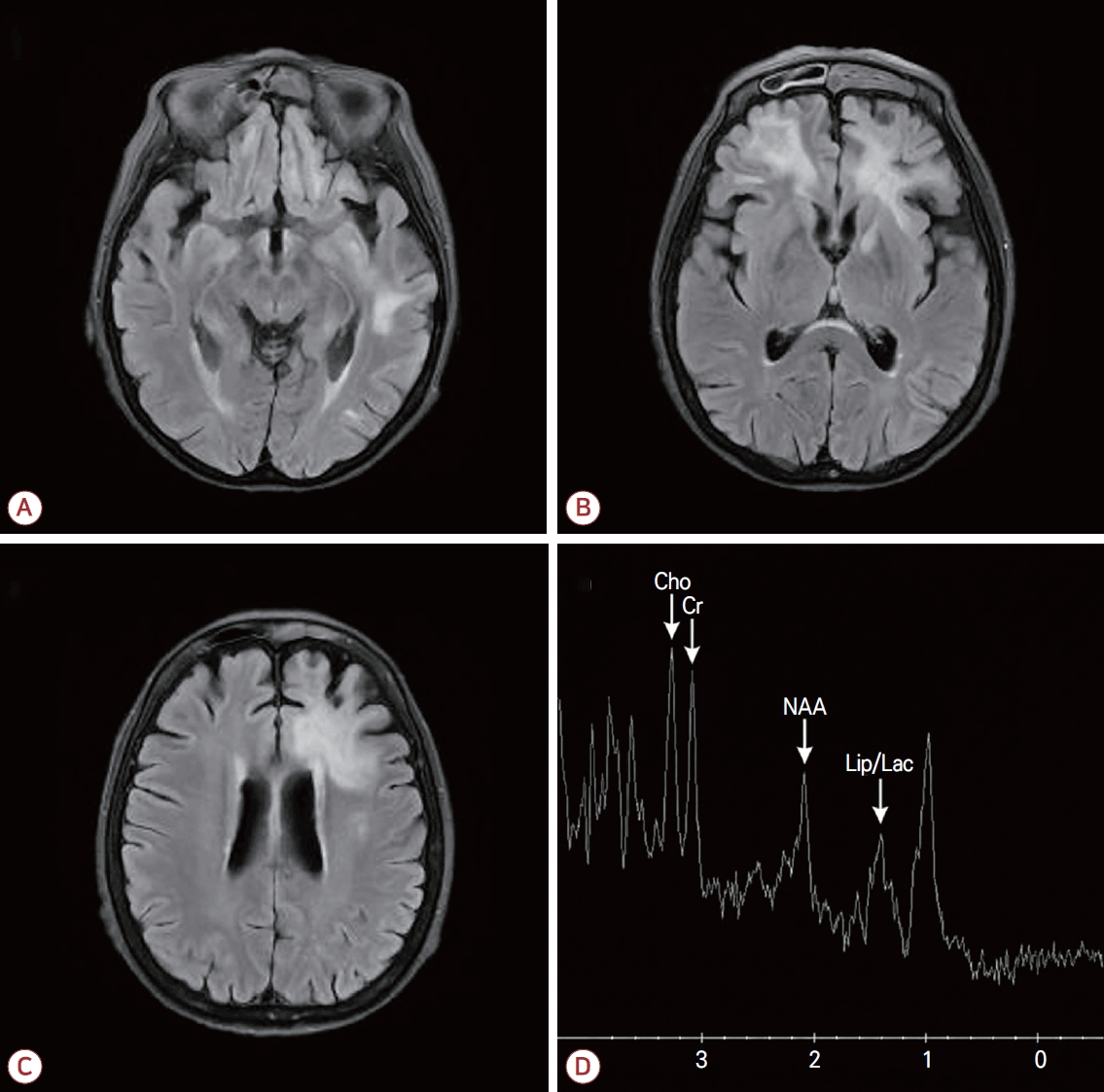
Follow-up brain magnetic resonance imaging and magnetic resonance spectroscopy findings. (A-C) FLAIR images obtained 1 month after the previous study reveal (A) a newly developed hyperintense lesion in the bilateral occipital subcortical regions and (B, C) increased extent of the pre-existing confluent hyperintense lesions in the bilateral frontal lobes. (D) Magnetic resonance spectroscopy obtained from the left frontal subcortical region demonstrates a markedly decreased N-acetylaspartate (NAA) peak, a slightly increased choline peak, and an increased lipid peak, findings consistent with demyelination. Cho; choline, Cr; creatine, Lip/Lac; lipid and lactate, FLAIR; fluid-attenuated inversion recovery.
혈액 검사에서 백혈구는 3,570/mm3였고 림프구가 24% (정상 범위, 28-39)로 감소되어 있었다. 림프구 아형 분석 결과 CD3+ T림프구는 89% (정상 범위, 56-86), CD3+CD4+ T림프구(도움 T림프구) 28% (정상 범위, 33-58), CD3+CD8+ T림프구(억제 T림프구) 62% (정상 범위, 13-39)로 CD4/CD8 비가 역전되었으며, 특히 CD19+ B림프구는 0% (정상 범위, 5-22)로 말초혈액에서 현저히 감소되었다. 혈청 면역글로불린(immunoglobulin, Ig)은 IgG 404 mg/dL (정상 범위, 870-1,700), IgA 44 mg/dL (정상 범위, 110-410), IgM <25 mg/dL (정상 범위, 46-260)로 저감마글로불린혈증이 확인되어 환자는 가슴샘종 병력과 부합하는 굿증후군(Good syndrome)으로 판단되었다.
감별 진단을 위하여 시행한 혈청 JCV에 대한 중합효소사슬 반응(polymerase chain reaction, PCR)은 음성이었으나 뇌척수액 JCV PCR 검사에서는 양성이 확인되었다. 중추신경계 자가면역질환 감별을 위한 혈청 신생물딸림항체, 항아쿠아포린-4항체, 항MOG항체, 류마티스인자, 항핵항체는 모두 음성이었으며 CSF1R유전자를 포함한 백질형성장애 관련 차세대염기서열분석에서도 병원성 변이는 발견되지 않았다. 뇌척수액 검사에서 백혈구 0/mm3, 단백질 25.7 mg/dL, 포도당 50 mg/dL (혈당 91 mg/dL)로 정상 범위였으며 세포학 검사에서 악성 세포는 관찰되지 않았다. IgG 지수는 0.77 (정상 범위, ≤ 0.7), 올리고클론띠는 1형이었다. 뇌척수액 단순포진바이러스(herpes simplex virus) 1형과 2형, 수두대상포진바이러스(varicella zoster virus), 엡스타인-바바이러스(Epstein-Barr virus), 거대세포바이러스(cytomegalovirus) PCR 및 14-3-3 단백질 검사는 모두 음성이었다. 흉부 및 복부컴퓨터단층촬영검사에서 가슴샘종의 잔존 또는 재발, 이외에 다른 암 질환을 시사하는 소견은 발견되지 않았다.
임상 소견과 영상 및 뇌척수액 검사 결과를 종합하여 굿증후군 환자에서 발생한 PML로 진단하였다. 확진을 위해서 뇌 생검을 고려하였으나 환자의 극심한 영양 결핍 상태와 인플루엔자균와 폐렴알균에 의한 폐렴, 칸디다 질염, 장염, 양측 신우신염 등 다발 감염이 병발하여 전신 마취의 위험도가 높아 시행하지 못하였다. 다발 감염에 대해 피페라실린/타조박탐 4.5 g을 8시간 간격으로 3일간 정맥 투여한 후 레보플록사신 500 mg 하루 1회, 메트로니다졸 500 mg을 하루 3회 정맥 및 경구 투여하였다. JCV PCR 결과 확인 전 중추신경계 탈수초질환과 감별을 위하여 고용량 메틸프레드니솔론(methylprednisolone) 1 g을 3일간 정맥 투여하였으나 신경학적 호전은 관찰되지 않았으며 보호자 요청에 따라 입원 7일째에 퇴원하였다. 입원 기간 동안 신경계 증상은 호전이나 악화 없이 유지되었으며 감염은 부분적으로 조절되었다. 이후 환자는 퇴원 약 1개월 후 사망하였다.
임상 소견과 영상 및 뇌척수액 검사 결과를 종합하여 굿증후군 환자에서 발생한 PML로 진단하였다. 확진을 위해서 뇌 생검을 고려하였으나 환자의 극심한 영양 결핍 상태와 인플루엔자균와 폐렴알균에 의한 폐렴, 칸디다 질염, 장염, 양측 신우신염 등 다발 감염이 병발하여 전신 마취의 위험도가 높아 시행하지 못하였다. 다발 감염에 대해 피페라실린/타조박탐 4.5 g을 8시간 간격으로 3일간 정맥 투여한 후 레보플록사신 500 mg 하루 1회, 메트로니다졸 500 mg을 하루 3회 정맥 및 경구 투여하였다. JCV PCR 결과 확인 전 중추신경계 탈수초질환과 감별을 위하여 고용량 메틸프레드니솔론(methylprednisolone) 1 g을 3일간 정맥 투여하였으나 신경학적 호전은 관찰되지 않았으며 보호자 요청에 따라 입원 7일째에 퇴원하였다. 입원 기간 동안 신경계 증상은 호전이나 악화 없이 유지되었으며 감염은 부분적으로 조절되었다. 이후 환자는 퇴원 약 1개월 후 사망하였다.
고 찰
본 증례와 같이 아급성으로 진행하는 신경계 증상과 광범위한 탈수초 병터를 보이는 경우 중추신경계 탈수초질환, 자가면역뇌염, 종양성 병터, PML 등을 감별 진단해야 한다. 중추신경계 탈수초질환이나 자가면역질환의 경우 면역 치료에 대한 반응 자체가 진단의 단서가 될 수 있다. 본 증례에서는 PML을 감별하기 위해서 뇌조직 검사를 고려하였으나 감염 및 전신 상태가 불량하여 시행하지 못하였으며, 대신 뇌척수액에서 JCV PCR을 확인하였다. 미국신경과학회에서 발표한 진단 기준에 따르면 뇌조직 검사에서 탈수초(demyelination), 기이 별아교세포(bizarre astrocytes), 희소돌기아교세포핵비대(enlarged oligodendrogilal nuclei)의 세 가지 특징 병리 소견이 관찰되고 면역조직화학 혹은 전자현미경 검사에서 JCV가 확인되거나 뇌조직 JCV PCR이 양성일 경우 PML로 진단할 수 있다[1]. 그러나 전신 상태가 불량하여 뇌조직 검사가 어려운 경우에는 전형적인 임상 경과와 영상 소견이 동반되고 뇌척수액에서 JCV PCR 양성이 확인되면 병리학적 확정 없이도 확실한(definite) PML로 진단할 수 있다[1].
PML은 주로 면역 저하 상태의 환자에서 발생하는 기회 감염 질환으로 HIV 감염, 혈액암, 면역억제제 사용 등이 주요 위험인자로 알려져 있다[2]. 본 증례에서는 혈액 검사에서 말초혈액의 B림프구의 현저한 감소와 CD4/CD8 T림프구 비율의 역전이 관찰되어 복합 면역결핍 상태를 시사하였다. 여기에 가슴샘종의 병력을 종합할 때 굿증후군에 의한 면역결핍 상태로 판단된다[3]. 굿증후군은 주로 40-70세에서 발병하며 유병률은 약 500,000-700,000명당 1명으로 보고되는 드문 질환이다. 전 세계적으로 보고된 증례는 400건 미만으로 주로 유럽과 아시아에서 보고되었다[4]. 병태생리학적으로는 골수에서 B림프구가 초기 전구 단계(pro-B cell)에서 분화가 차단되어 성숙 B세포가 결핍되고, 이로 인하여 저감마글로불린혈증이 발생한다. 이러한 변화는 가슴샘종에 의한 골수 미세환경 교란, 면역조절인자의 불균형, 후천 조절 이상 등이 복합적으로 작용한 결과로 추정된다. 또한 T세포 면역 이상 역시 중요한 특징으로 가슴샘 구조 파괴와 음성선택장애로 인한 조절 T림프구 감소, 자가반응 T림프구의 말초 유출 등이 관여하는 것으로 알려져 있다. 이로 인해 세포 및 체액 면역기능 모두 저하되어 기회 감염의 위험이 증가한다. 상·하기도부터 위장관, 비뇨기, 피부 및 연조직, 골관절, 점막에 이르기까지 다양한 부위에서 감염을 일으킬 수 있으나 특히 순환기계 감염이나 중추신경계 침범은 예후를 악화시키는 주요 요인이다[5].
현재까지 굿증후군에서 PML의 발병은 5예가 보고되었다(Table). 보고된 증례들에서 가슴샘종 절제술 후 PML이 발생하기까지의 간격은 약 5개월에서 8년까지 다양하였으며 일부 환자에서는 수술 치료를 시행하지 못하였다. 임상적으로는 빠르게 진행하는 신경계 증상을 보였고 혈액 검사에서는 CD4/CD8 비 역전과 저감마글로불린혈증이 관찰되었다. 또한 한 증례를 제외하고는 대부분 수개월 이내에 사망하는 불량한 예후를 보였다. PML 치료로 스테로이드, 면역글로불린 정맥 주사(intravenous immunoglobin, IVIG), 시도포비어(cidofovir), 메플로퀸(mefloquine) 등의 다양한 약제가 시도되어 왔으며 최근에는 면역검문억제제(immune checkpoint inhibitors)를 적용한 증례 보고도 발표되고 있다. 그러나 현재까지 이러한 치료법들의 효과를 명확히 입증하는 충분한 근거는 부족하여 표준 치료로 확립된 약제는 없다. 굿증후군에서 IVIG가 저감마글로불린혈증을 교정하고 감염 발생을 감소시키는 데 효과적이지만 세포매개 면역결핍을 동반하는 복합 면역결핍 상태로, IVIG만으로는 면역기능을 회복시키는 데에는 다소 제한이 있다. 본 증례에서는 다발 감염 및 전신 상태 악화, 보호자의 요청으로 인하여 고용량의 스테로이드 투여 외에 추가적인 면역 치료를 시행하지는 못하였다.

Previous reported cases of progressive multifocal leukoencephalopathy
본 증례는 가슴샘종 절제술 이후 11년이 경과한 환자에서 굿증후군에 의한 면역결핍 상태와 이에 따른 PML이 발생한 사례이다. 뇌조직 검사는 시행하지 못하였으나 임상 경과와 영상 소견, 뇌척수액 JCV PCR 양성을 바탕으로 PML로 진단할 수 있었다. 이는 가슴샘종 절제 이후 장기간이 경과한 환자에서도 굿증후군과 기회 감염의 위험이 지속될 수 있음을 시사하며 가슴샘종 절제 환자에서 장기적인 면역 상태 평가와 추적 관찰이 필요함을 시사한다.