뇌 아밀로이드 혈관병증-관련염증의 자기공명영상 소견
Magnetic Resonance Image Findings of Cerebral Amyloid Angiopathy Related Inflammation
Article information
66세 여자가 2개월 전부터 진행하는 이상행동과 보행장애로 내원하였다. 신경학적 진찰에서 오른쪽 반신불완전마비로 보행이 불가능하였고 뇌신경기능장애나 감각신경 이상은 없었다. 언어의 유창성이 저하되었고 이름 대기에 어려움을 호소하였다. 뇌 magnetic resonance imaging (MRI)의 액체감쇠역전회복영상(fluid-attenuated inversion recovery imaging)에서 양쪽 이마엽과 왼쪽 마루관자후두엽의 광범위한 혈관성 부종이 보였고 조영증강은 없었다. 감수율강조영상(susceptibility weighted image)에서 피질하 지역의 다발적인 저신호강도 병변이 관찰되어 뇌 아밀로이드 혈관병증-관련염증(cerebral amyloid angiopathy related inflammation, CAA-RI)이 의심되었고(Fig. A-C), F-18 플로르베타벤 뇌 양전자방출단층촬영(F-18 florbetaben positron emission tomography)을 통하여 양측 피질하 지역의 전반적인 아밀로이드 침착을 확인하였다. 덱사메타손 치료 후 근력저하와 기억력저하가 호전되었고, 치료 2주와 1년째 추적한 뇌 MRI에서 혈관성 부종이 감소하였다(Fig. D-I). CAA-RI은 드문 질환으로 이상행동, 인지기능 저하, 뇌전증 및 국소신경학적 결손으로 나타나며, 혈관벽에 있는 베타아밀로이드의 염증 반응이 원인으로 생각된다[1]. 주로 비대칭적으로 발생하며 면역 치료에 반응이 좋다[2].
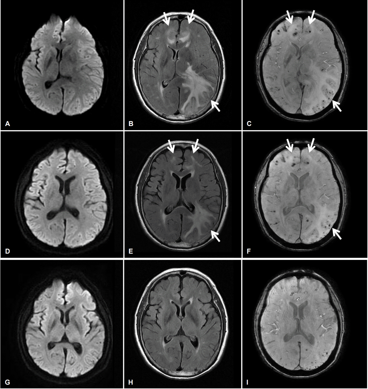
Serial diffusion weighted images (DWI), fluid attenuated inversion recovery (FLAIR) images and susceptibility weighted images (SWI) of patient. (A) In initial DWI reveals no abnormal signal intensity, but in FLAIR image shows high signal intensity lesions with edema on bilateral frontal and left parieto-temporo-occipital lobes (arrows) (B). (C) SWI reveal subcortical microbleeds near the abnormal FLAIR signal intensity lesions (arrows). (D-F) Immediate follow up images after treatment with dexamethasone for 2 weeks shows much improvement of previous abnormal lesions in T2 FLAIR image (E, arrows) but the microbleeds on SWI are persisted (F, arrows). (G-I) One year later in follow up magnetic resonance imaging, the T2 FLAIR abnormal signal intensity lesions are disappeared (H).