소리조짐이 동반된 뇌전증으로 발현한 뇌이소증
Cerebral Heterotopia Presenting as Epilepsy with Auditory Aura
Article information
J Korean Neurol Assoc. 2018;36(4):411-412
Publication date (electronic) : November 1, 2018
doi :
http://dx.doi.org/10.17340/jkna.2018.4.35
received : May 27, 2018 , rev-recd : June 26, 2018 , accepted : June 26, 2018 .
37세 여성이 전화통화 중 좌측 귀에서 윙윙거리는 소리가 들리고 전신이 뻣뻣해지며 3-4분간 의식소실 후 30분 동안 혼돈증상이 지속되어 입원하였다. 병력에서 1년 전 유사 증상이 있었다. 뇌 자기공명영상에서 우측 가쪽 뇌실의 뒤통수뿔에 이소증이 확인되었다(Fig.). 24시간 비디오뇌파검사에서는 특이 소견은 관찰되지 않았지만 뇌전증 정의에 따라 뇌전증을 진단하고 항뇌전증약을 투약하였다. 이소증은 무증상부터 조절되지 않는 뇌전증까지 다양한 임상양상을 나타낸다[1]. 편측 소리조짐은 반대편 측두 신피질로 국소화될 수 있으나 환자가 소리의 방향이나 내용을 구분하기 어려운 경우가 많아 국소화시키기 어려울 때가 많다[2]. 전화통화 중 소리조짐이 발생하였고 뇌병변은 우측에서 이소증이 확인된 드문 사례를 보고하는 바이다.
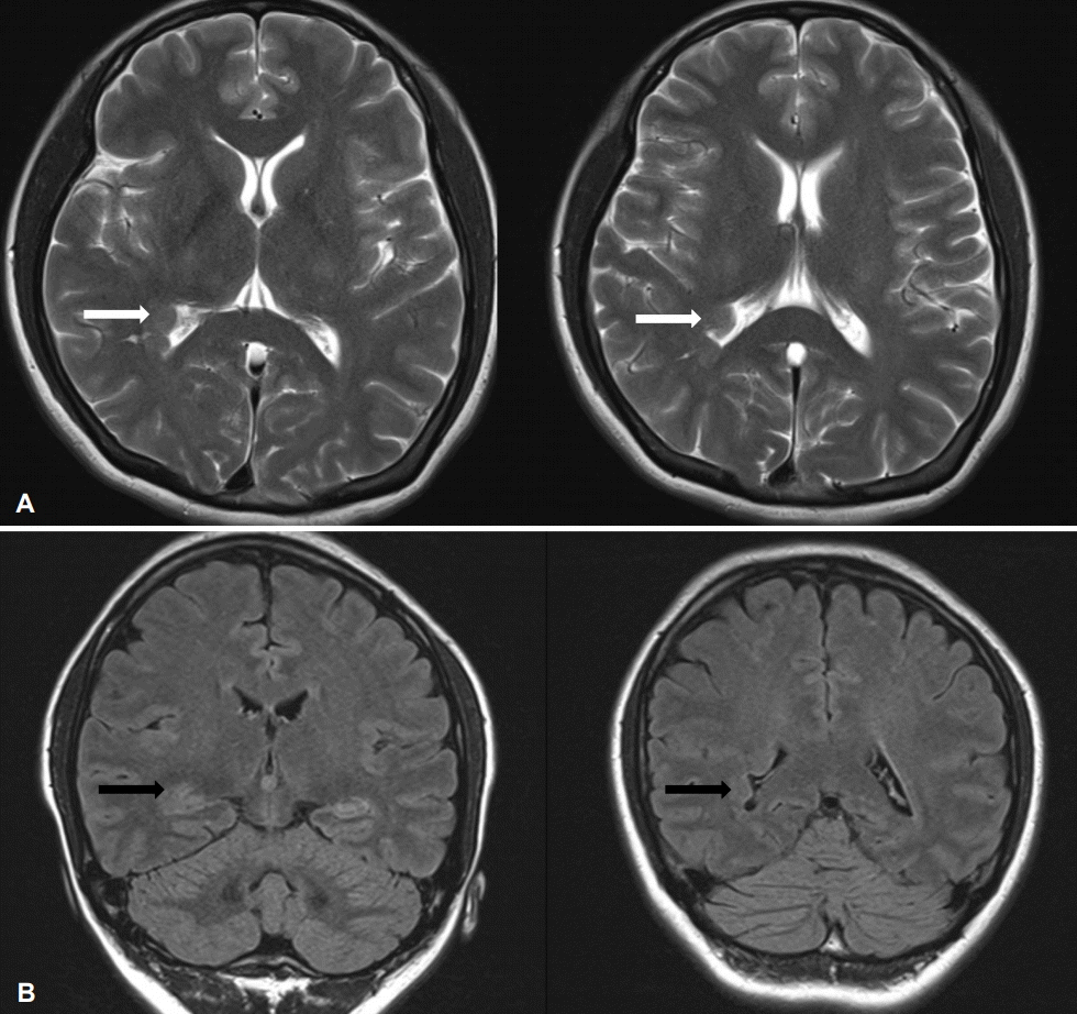
T2 weighted axial images show smooth nodular lesion on occipital horn of right lateral ventricle and it is isointense to grey matter (A, white arrows). Coronal images of fluid attenuated inversion recovery (FLAIR) reveal nodular lesion with indistinct margin on subependymal regions of right lateral ventricle (B, black arrows).
References
1. Barkovich AJ, Kuzniecky RI. Gray matter heterotopia. Neurology 2000;55:1603–1608.
2. Florindo I, Bisulli F, Pittau F, Naldi I, Striano P, Striano S, et al. Lateralizing value of the auditory aura in partial seizures. Epilepsia 2006;47 Suppl 5:68–72.
Article information Continued
Copyright © 2018 Korean Neurological Association