전립선암에 의한 경질막 전이
Dural Metastases from Prostate Cancer
Article information
70세 남자가 2009년에 전립선암으로 진단되었고, 이후 척추 및 다발골전이 보여 약물을 변경하면서 8차례 항암 치료하였다. 2개월 전부터 눈이 침침하였고, 안과검진상 시신경유두부종이 보였다. 뇌 자기공명영상상 뇌수막이 전반적으로 조영증강을 보이면서 두꺼워져 있었고, 국소결절을 보였다(Fig. A). 자기공명정맥조영술에서는 경질막비후에 의해 위시상정맥동의 전반적인 협착이 보여(Fig. B) 암세포의 침착이 의심되었다. 우측 전측두부에서 뇌수막과 결절을 절제하였다(Fig. C). 병리검사상 경질막을 침범하는 꽈리샘암종(acinar adenocarcinoma, gleason score: 8)이 보였고(Fig. D, E), 이는 전립선특이항원염색에 잘 염색되어(Fig. F) 전이전립선암으로 확진하였다.
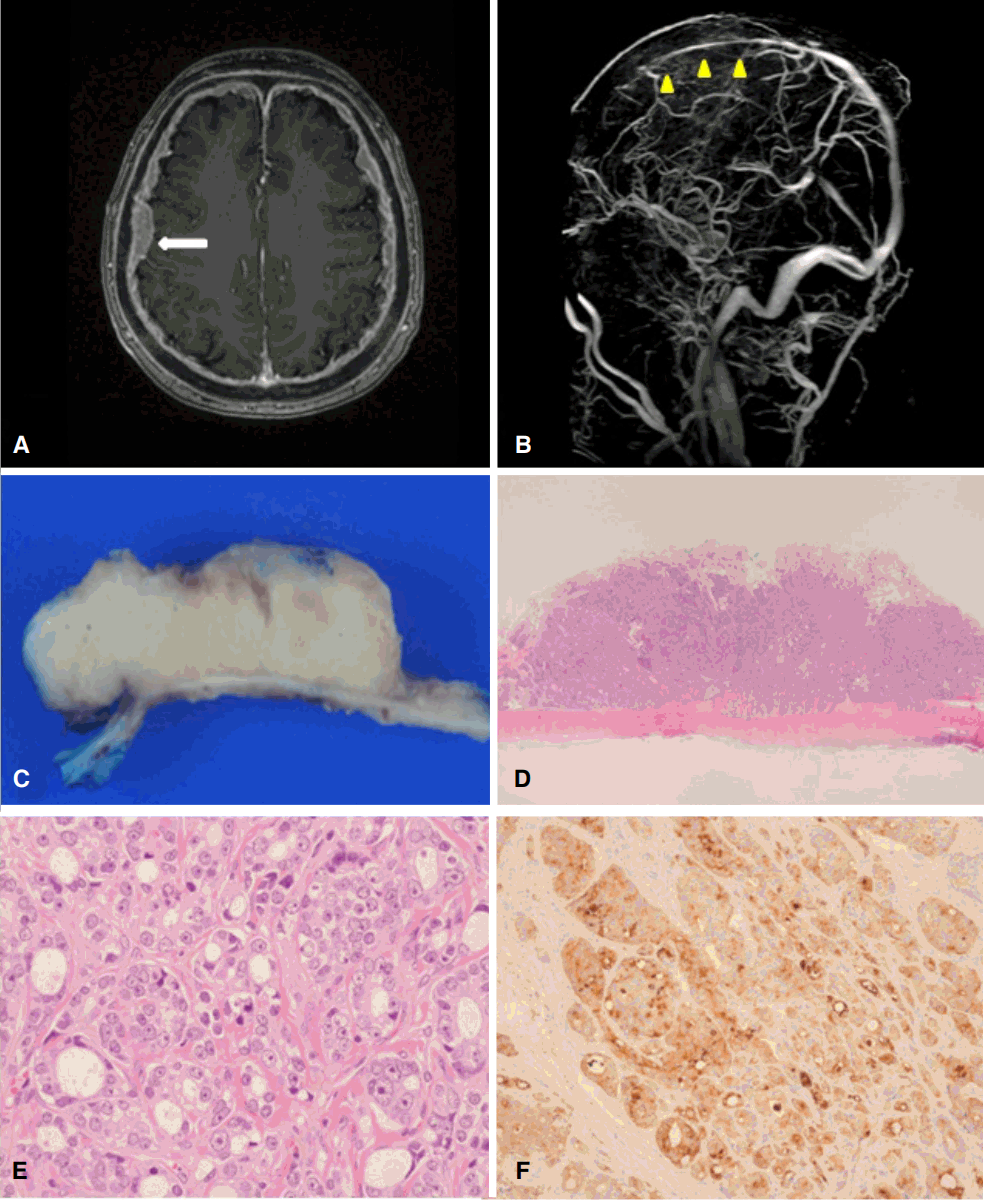
Brain images and pathologic findings of patient. T1-weighted enhanced image (A) shows diffuse thickening and enhancement of pachymeninges (white arrow: nodular mass lesion). Magnetic resonance venography (B) shows diffuse stenosis of the superior sagittal sinus, which appears to be caused by infiltration of tumor cells (yellow triangles). Gross photo (C) shows a nodular lesion which its cut surface is yellowish white, firm and granular involves pachymeninges. Microscopic photo (D) shows a tumor nodule involving underlying meninges is observed in low power view (H&E, ×10). Microscopic photo (E) shows fused tumor glands which are graded Gleason 4 are observed. Tumor cells have large nuclei with prominent nucleoli in high power view (H&E, ×400). Microscopic photo (F) shows immunohistochemical staining of specimens with prostate specific antigen. Tumor cells show diffuse immunopositivity for prostate-specific antigen (×400).
전립선암의 경질막 전이는 드물어서, 현재까지 산발적인 보고만 있을 뿐이며, 한 단일병원기반 연구에 의하면 6,282명의 전이전립선암에서 28명(0.4%)만이 확인되었다[1]. 전립선암의 뇌내 전이는 드물지만 경질막이 가장 흔한 부위이다[2]. 전립선암의 치료 방법이 다양해지면서 기대여명이 길어지므로, 유병률은 늘어날 가능성이 있다.