신경혈관압박이 확인된 상사근 근잔떨림
Superior Oblique Myokymia Associated with Neurovascular Cross Compression
Article information
Trans Abstract
Superior oblique myokymia (SOM) is a rare disorder characterized by unilateral paroxysmal oscillopsia or diplopia. Recent studies revealed that SOM can be associated with neuro-vascular cross compression (NVCC) of the trunk of the trochlear nerve. Although it frequently occurs without any underlying systemic disease or concurrent neurologic sign, we need to consider this NVCC especially in cases with persistent disturbing symptoms. Hereby, we present two cases of SOM whose neuroimaging studies suggest NVCCs and, discuss recent update of the pathomechanism of SOM.
상사근 근잔떨림(superior oblique myokymia)은 단안에서 발생하는 상사근의 돌발(paroxysmal)불수의 흥분으로 인해 발생하며, 하방주시에 자발적인 상사근의 진동으로 회선, 수직진동시각(oscillopsia)이나 복시를 초래하는 드문 질환이다[1]. 1906년 Duane이 상사근을 포함하는 단안의 회선안진을 최초로 보고하였으며, 1970년 Hoyt와 Keane이 간헐진동시를 보이는 상사근의 비정상운동에 대해 ‘상사근 근잔떨림’이라고 처음 명명하였다[2]. 스트레스나 수면부족, 알코올 등에 의해 유발될 수 있고, 증상은 대개 수십 초 정도 지속되며, 하루에도 수초에서 수분 간격으로 여러 차례 재발하는 경향을 보인다. 특히 책을 읽거나 전화기 사용 등 하방주시에서 증상이 악화된다. 대부분 양성 질환이나 다발성 경화증이나 후두개와종양 등 구조적 뇌병터가 있는 경우에도 발생할 수 있으며, 반얼굴연축(hemifacial spasm)이나 삼차신경통과 같은 신경혈관압박(neurovascular cross compression)에 의해서도 나타날 수 있다[3].
저자들은 복시와 진동시를 주증상으로 방문하여 비디오안구운동검사로 상사근 근잔떨림으로 진단한 두 증례를 확인하여 문헌고찰과 함께 보고하고자 한다.
증 례
증례1
45세 여자가 약 5년 전부터 시작된 간헐복시로 내원하였다. 어지럼과 진동시를 동반하고 있었고, 피곤하거나 집중할 경우 악화되었으며, 특히 신문을 보거나 핸드폰을 사용하는 등 하방주시에서 발생하였다. 최근에는 빈도가 증가하여 하루에 수십 차례 증상이 나타났으며, 심한 경우 며칠 동안 지속되었다고 하였다. 과거력과 가족력에서 특이사항은 없었으며, 흡연, 음주는 하지 않았고 두부외상 및 약물복용력은 없었다. 활력징후는 혈압 122/68 mmHg, 맥박 84회/분, 체온 36.6℃였다.
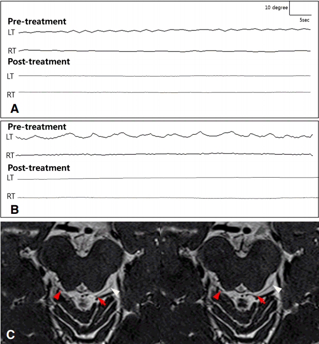
신경학적 진찰에서 의식은 명료하였고, 인지기능은 정상이었다. 동공의 크기 및 빛반사는 정상이었으며 안구운동 범위도 정상이었으나 전방과 하방주시 시 좌안에서 회선눈떨림이 관찰되었다. 근력과 감각기능, 소뇌기능검사는 정상이었으며, 심부건반사, 바빈스키징후검사에서도 이상 소견은 보이지 않았다. 일반 혈액검사와 당화혈색소, 콜레스테롤을 포함한 일반 생화학검사에서 정상이었으며, 갑상선기능검사에서도 이상 소견을 보이지 않았다. 비디오안구운동검사에서 좌안에서 회선방향의 근잔떨림이 확인되었으며(Fig. A upper panel, Video 1), 안와 자기공명영상에서 안구주변이나 뇌간부, 소뇌를 포함한 뇌실질에 음영변화나 조영증강 등의 이상 소견은 보이지 않았으나, 재구성한 3D sampling perfection with application-optimized contrasts using different flip angle evolution (SPACE)영상에서 좌측 도르래신경이 상소뇌동맥가지로부터 압박됨을 확인할 수 있었다. 상사근 근잔떨림으로 진단하고 옥스카바제핀(oxcarbazepine) 300 mg 하루 한 번으로 투약을 시작하였다. 증상은 초기에 약간 호전되었으나 2주 후 추적 관찰시 다시 복시와 진동시를 호소하여 옥스카바제핀을 300 mg 하루 두 번으로 증량한 후 증상이 호전되었다. 한 달 후 추적 관찰에서 진동시나 복시 등의 증상은 없었으며, 재시행한 비디오안구운동검사에서 눈떨림도 관찰되지 않았다(Fig. A lower panel).
증례2
33세 남자가 2년 전부터 간헐적으로 눈이 떨리며 진동시로 내원하였다. 증상이 생길 때마다 어지럼, 좌측 두정부 부위의 경미한 두통이 동반되었고, 뚜렷한 복시는 호소하지 않았으며, 시력감소도 없었다. 눈떨림과 진동시는 피곤할 때 악화되어 5초 정도 지속되었으며, 하루에 7-8회 반복된다 하였다. 과거력에서 두부외상이나 뇌혈관질환 등의 병력은 없었으며 가족력 및 약물복용력에서도 특이사항은 없었다. 주 3회 소주 3병 정도의 음주력이 있었으며, 활력징후는 혈압 135/84 mmHg, 맥박 70회/분, 체온 36.7℃였다.
신경학적 진찰에서 의식은 명료하였고, 뇌신경검사에서 안구운동범위 및 동공의 크기와 반응, 안압은 정상이었으나 좌안에서 지속적인 회선눈떨림이 관찰되었다. 양측에서 근력 및 감각저하는 없었고, 심부건반사나 병적반사는 관찰되지 않았으며, 소뇌기능검사에서도 특이 소견은 보이지 않았다. 일반 혈액검사와 일반 생화학검사는 정상이었고, 갑상선기능 및 항체, 비타민 B12, 지질단백질, 자가면역질환 항체를 포함한 검사실검사에서 정상 소견을 보였으며, 비디오안구운동검사에서 좌안의 지속회선안구운동이 관찰되었다(Fig. B upper panel, Video 2). 뇌 자기공명영상에서 안와, 해면정맥동을 포함한 뇌실질에 특이 소견을 보이지 않았으나 재구성한 3D SPACE영상에서 좌측 도르래신경이 상소뇌동맥가지로부터 압박됨을 확인할 수 있었다(Fig. C). 상사근 근잔떨림으로 진단한 후 옥스카바제핀 300 mg 하루 2회로 투약을 시작하였고, 유의한 호전 소견을 보이지 않아 450 mg 하루 2회로 증량하였고, 2주 후 증상은 소실되어 현재까지 유지 중이다.

Video-oculography (VOG) recordings of the patients. (A) VOG recordings of Patient 1 (45-year-old woman) revealed torsional oscillation on the left eye during primary gaze (upper panel). After oral oxcarbazepine 300 mg twice a day for a week, ocular movements were subsided (lower panel). (B) VOG recordings of Patient 2 (31-year-old man) also showed torsional oscillation on the left eye during primary gaze (upper panel). These ocular movements were subsided after a treatment with oral oxcarbazepine 450 mg twice a day (lower panel). (C) Axial high-resolution 3D heavily T2-weighted images using SPACE (sampling perfection with application-optimized contrasts using different flip angle evolution) of Patient 2 showed contact between the medial superior cerebellar artery branch and the cisternal segment of left trochlear nerve 1.5 mm distal to the point of exit from the brainstem (red arrow). Note that the transition zone of left trochlear nerve is barely visible and thinned, while the more distal portion of the left trochlear nerve (white arrowhead) and the transition zone of right trochlear nerve (red arrowhead) have a normal thickness.
고 찰
상사근 근잔떨림은 환자가 단안의 진동시, 수직 혹은 회선복시 등의 증상을 호소할 때 신경학적 진찰과 비디오안구운동검사를 통해 비교적 쉽게 진단이 가능하고, 약물 치료에도 효과적이다. 대부분 동반된 전신질환이나 신경계 증상 없이 나타나지만 경미한 두부외상이나 뇌간경색, 동정맥루, 4번신경마비나 4번신경의 별아교세포종(astrocytoma) 또는 신경압박이나 다발성 경화증과 같은 뇌내 병터에 의해서도 발생할 수 있기 때문에 뇌 자기공명영상 등으로 구조적 이상을 확인하는 것이 필요하다[1].
상사근 근잔떨림의 기전에 대해서는 확실하지는 않지만, 상사근의 비정상폭주에 의해 근잔떨림이 발생하는 것으로 생각되며, 도르래신경(trochlear neve)의 신경막역치의 병리적 변화가 관여된다[4]. 최근의 연구에 의하면 삼차신경통이나 반얼굴연축과 같이 신경혈관압박에 의해 발생할 수 있으며[5], 증상이 지속되는 경우에는 수술을 통한 미세혈관감압술을 통해 증상의 호전과 치료를 보고한 바 있다. Samii 등은 중뇌로부터 나온 우측 도르래신경이 동맥, 정맥으로 인해 압박되어 발생한 상사근 근잔떨림 환자에서 미세혈관감압술을 통해 안구움직임이 호전되어, 22개월간의 추적 관찰에서 재발하지 않았다고 보고하였으며[6], Mikami 등은 약제 치료에서 효과가 없던 환자에서 상소뇌동맥가지로 인한 도르래신경의 압박을 수술로써 해소하였다[7]. 또한 Hashimoto 등[8]은 간헐진동시로 내원한 50세 여자 환자에서 자기공명영상을 통해 후대뇌동맥의 분지인후맥락동맥(posterior choroidal artery)에 의한 도르래신경압박을 보고하였으며, Yousry 등[9]은 시력저하와 복시로 온 6명의 우안 상사근 근잔떨림 환자에서 상소뇌동맥에 의한 도르래신경 뿌리의 직접적 신경압박을 자기공명영상을 통해 보고한 바 있다. 본 증례에서도 재구성한 뇌 자기공명영상에서 미세신경혈관압박이 의심되는 소견이 관찰되었으며, 이는 상사근 근잔떨림의 병태생리로, 신경혈관압박이 원인일 수 있음을 추정하는 근거로 이에 관한 체계적인 임상 연구가 필요하겠다.
치료는 기질적인 원인이 감별되고 환자가 증상에 대한 자각이 없을 경우 경과 관찰로 충분하나, 복시나 진동시에 대해 불편감을 호소하거나 일상생활에 문제가 생길 경우 약물 치료를 시작하며, 호전이 없을 경우 수술 치료도 고려할 수 있다[1]. 약물 치료는 도르래신경의 병적 흥분의 빈도를 감소시켜 증상을 완화시키는 막안정제로써 도르래운동신경을 억제시켜주는 카르바마제핀(carbamazepine)이 가장 흔하게 사용되는데[4], 카르바마제핀의 경우 백혈구감소증, 급성 신부전, 혈전색전증, 부정맥 등의 심각한 부작용을 일으킬 수 있으므로 면밀한 관찰이 필요하다[1]. 수술 치료는 증상이 심하거나 약물투여로도 효과가 없는 경우 상사근 힘줄절단술(tenotomy)이나 동측 하사근 절제술로써 진동시의 호전을 보일 수 있지만, 수술 후 의인도르래신경마비로 인한 이차하방 복시와 상사시가 약 35%의 환자에서 발생하였다고 하였다[10]. 또한 신경혈관압박이 확인된 경우 도르래신경의 감압술을 시행하여 증상의 호전을 기대할 수 있겠다[6].
상사근 근잔떨림은 드문 질환이지만 특징적 안구운동을 보이기 때문에 신체진찰이나 비디오안구운동검사 등을 통해 쉽게 진단이 가능하고, 대부분 동반질환 없이 발생한다. 그러나 본 증례와 같이 상사근 근잔떨림 환자의 뇌 자기공명영상에서 신경혈관압박이 관찰되며 이러한 환자에서 경구약제에 반응하지 않는 경우 감압술 등의 적극적 치료를 고려할 수 있겠다.
Supplementary Material
Video 1.
Video Frenzel's goggles of Patient 1 showed continuous torsional oscillation in the left eye and notable improvement of micro-tremor when we started oxcarbazepine 300 mg twice a day on a subsequent video.
Video 2.
Frenzel's goggles of Patient 2 showed continuous torsional oscillation in the left eye.