코로나바이러스감염증-19와 관련된 급성 괴사뇌병증
Acute Necrotizing Encephalopathy Associated with COVID-19
Article information
64세 여자가 내원 당일에 시작된 어지럼증과 구음장애로 응급실에 방문하였다. 환자는 의식이 빠르게 악화되면서 6시간째 혼수가 되고 뇌간반사가 소실되었다. 뇌 magnetic resonance imaging에서 뇌줄기와 양쪽 시상 및 안쪽 측두엽 병터가 보였고(Fig.), 부종과 주변부 확산 제한이 동반됐다. 뇌 computed tomography 혈관조영술은 정상이었다. 뇌척수액 검사에서 단백질이 222 mg/dL로 상승되었지만 백혈구(1/mm3)와 포도당(81 mg/dL)은 정상이었다. 환자는 내원 2일 전부터 기침 등 가벼운 상기도감염 증상이 있었고 이에 코로나바이러스감염증-19 감염을 의심하였다. Severe acute respiratory syndrome-coronavirus-2 (SARS-CoV-2) 비인두도말신속항원 검사와 중합효소사슬반응(polymerase chain reaction, PCR) 검사가 양성이었고, 그 외 단순포진바이러스, 대상포진바이러스, 엡스타인-바바이러스, 거대세포바이러스, 엔테로바이러스, 일본뇌염바이러스, 인플루엔자를 포함한 바이러스와 세균 배양 및 마이코플라즈마, 리스테리아, 보렐리아 등 세균 항체와 PCR 검사 그리고 결핵, 곰팡이, 매독, 기생충 검사는 모두 음성이었다. 비감염염증질환으로 신생물딸림(paraneoplastic)항체, 수초희소돌기아교세포당단백(myelin oligodendrocyte glycoprotein)과 아쿠아포린-4 (aquaporin-4)항체, 올리고클론띠, 글루탐산탈탄산효소(glutamic acid decarboxylase)항체, 갑상샘자가항체, 항핵항체, 항중성구세포질항체를 포함한 혈관염 검사들도 모두 음성이었다. 코로나바이러스감염증-19에 의한 급성 괴사뇌병증(acute necrotizing encephalopathy) 진단으로 렘데시비르, 메틸프레드니솔론과 면역글로불린 정맥 주사를 투약하였지만 호전이 없었고, 12일째 대량의 소장 출혈로 환자는 사망하였다. 급성 괴사뇌병증은 인플루엔자바이러스가 대표적인 원인이지만 SARS-CoV-2 감염도 보고되었다[1]. 병터는 뇌간과 양쪽 시상을 대칭적으로 침범하고, 출혈과 중심 괴사가 동반될 수 있다. 주변부의 고리 모양의 확산 제한이 급성 괴사뇌병증에서 특징적이며[2], 뇌척수액세포증가증이 없는 점에서 뇌염과 구분된다.
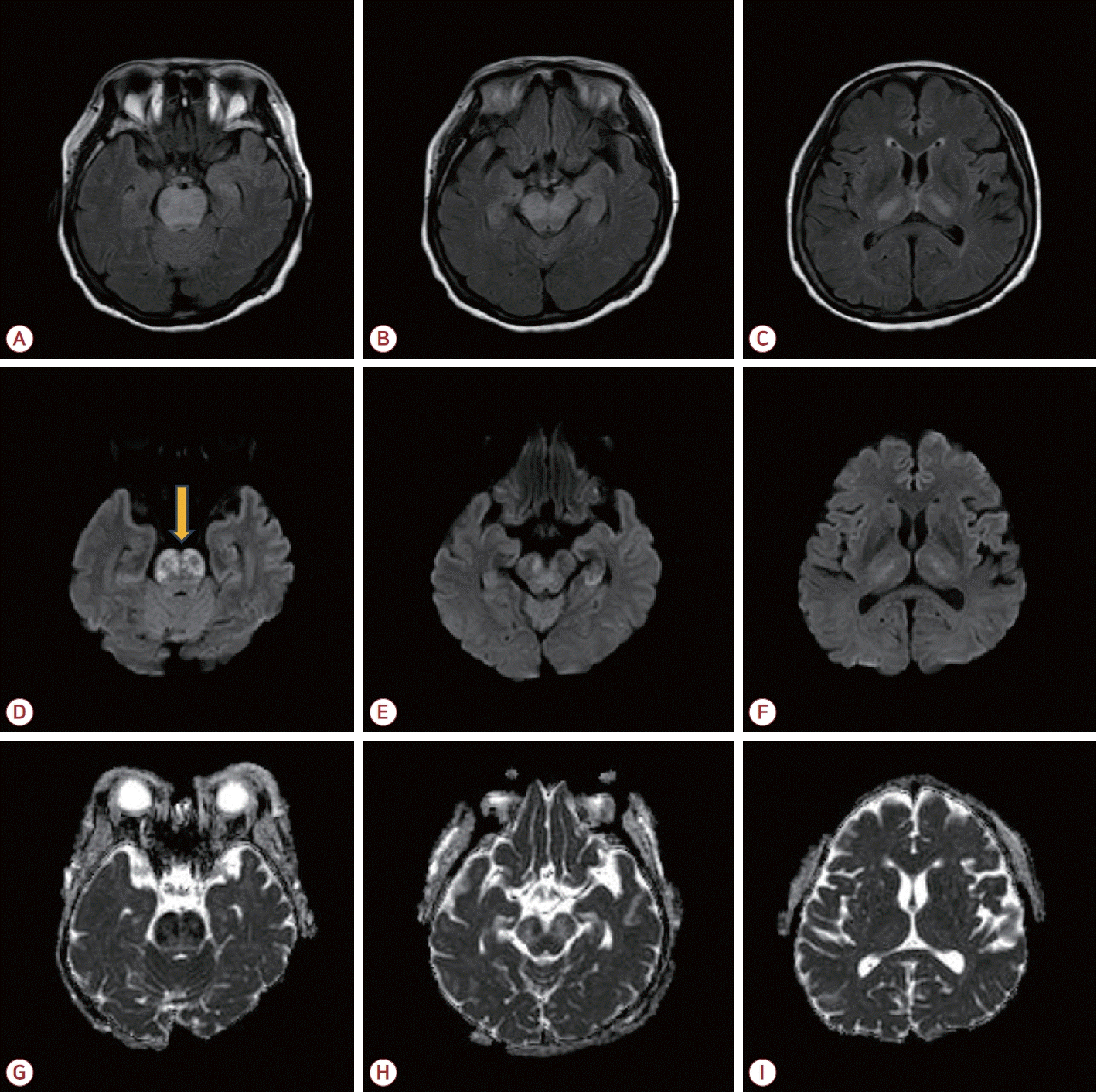
Brain MRI. (A-C) FLAIR images showed swelling and hyperintensities symmetrically involving the brainstem, bilateral medial temporal lobes, and thalami. (D-F) Diffusion-weighted imaging demonstrated diffusion restriction. Ring-shaped lesions with peripheral diffusion restriction (arrow in D) were noted in the pons. (G-I) Apparent diffusion coefficient images showed heterogeneous signal intensities suggesting coexistence of cytotoxic and vasogenic edema. MRI; magnetic resonance imaging, FLAIR; fluid attenuated inversion recovery.