반복되는 상지 심부정맥혈전증을 동반한 급성뇌경색의 원인
The Etiology of Acute Stroke with Experience of Repeated Upper Extremity Deep Vein Thrombosis
Article information
Trans Abstract
In stroke patients, upper extremity deep vein thrombosis (UEDVT) is uncommon compared with lower extremity. Unlike the blood stasis in lower extremity, UEDVT has been developed by secondary cause. We reported a case of stroke patient with repeated UEDVT, presenting superficial venous congestion, who was finally diagnosed with pulmonary adenocarcinoma. The cause of stroke was non-bacterial thromboembolism formed at the mitral valve. Our case shows that unexpected UEDVT should be closely evaluated for higher coagulable status such as a malignancy.
뇌졸중 환자에서 심부정맥혈전증은 잘 알려진 합병증 중 하나다[1]. 움직임이 제한된 환자의 하지에서 주로 발생하며, 운동기능 저하에 따른 혈액 저류로 설명된다[2]. 하지만 상지에서 심부정맥혈전증이 발생하는 경우는 매우 드물다.
상지에서 발생한 심부정맥혈전증 은 일반적으 로 대부분 이차적인 원인을 가지고 있다. 중심정맥 카테터 사용, 정맥영양, 심박동기 삽입이나 혈전이 발생하기 쉬운 조건(prothrombotic condition)에서 드물지 않게 발생하는 것으로 알려져 있으며, 폐색전증도 발생할 수 있다[3,4]. 따라서 상지심부정맥혈전증이 있으면 원인에 대한 평가가 필요하다.
본 증례에서는 상지피부정맥의 확장을 동반한 피부 병변을 보이는 뇌경색 환자에서 팔머리정맥혈전증을 진단하였고, 반복되는 혈전 발생 원인으로서 최종적으로 폐샘암종을 진단했던 환자에 대해 보고한다.
증 례
57세 남자가 왼손 근력저하(Medical Research Council[MRC] 척도 4), 저린 감각을 주소로 응급실을 내원하였다. 과거력으로 당뇨병, 심근경색이 있었고, 3개월 전 우측 팔머리정맥의 혈전증이 진단되어 리바록사반(rivaroxaban) 15 mg을 일 2회 3주 복용하였고, 이후 일 1회 20 mg으로 총 12주동안 복용 중이었다. 신경계진찰에서 좌측 상지 근력 저하와 감각 저하로 National Institute of Health (NIH) 뇌졸중척도 2점이 확인되었다. 뇌 컴퓨터단층촬영(computed tomography, CT)에서 저명한 저음영 소견은 없었고, 뇌 CT 혈관조영에서도 대뇌동맥 폐색은 관찰되지 않았다. 뇌 자기공명영상(magnetic resonance imaging, MRI)에서 양측 중대뇌동맥 영역의 다발허혈 병변이 확인되어(Fig. 1) 아스피린 100 mg, 클로피도그렐(clopidogrel) 75 mg, 로수바스타틴(rosuvastatin) 20 mg을 일 1회 복용하기 시작하였다. 입원 10일째 식도경유심초음파검사에서 승모판 부위 덩이(0.44×0.45 cm)가 발견되었다(Fig. 2-D). 활력징후에서 발열은 없었고 혈액검사에서 혈색소 16.2 g/dL, 백혈구 10,890/μL, 혈소판 136,000/μL, C-반응단백질(C-reactive protein) 0.71 mg/dL, 적혈구침강속도(erythrocyte sedimentation rate) 12 mm/hr로 감염심내막염을 의심할 만한 소견은 없었다. 4차례의 혈액배양검사에서 모두 음성이었다. 그러나 Duke 진단기준에 따라 가능성 있는(possible) 감염심내막염의 진단기준에 부합하여 경험적 항생제로 암피실린-설박탐(ampicillin-sulbactam) 4.5 g과 젠타마이신(gentamicin) 210 mg 일 1회 정맥주사 치료를 시작하였다. 입원 15일째 갑작스럽게 좌측 쇄골하 부위(subclavian area)의 통증과 부종을 호소하였으며, 점차 붓기는 왼손과 왼팔로 진행되었고 피부에는 정맥 울혈이 발견되었다(Fig. 2-A). 혈액응고검사에서 D-이합체(D-dimer) 2,439 ng/mL, 프로트롬빈시간(prothrombin time) 13.9 seconds, 국제표준화비율(international normalized ratio, INR) 1.20, C-단백항원 80.9%, C-단백활성도 89%, S-단백 항원 80.2%, S-단백활성도 65%, 항인지질항체(anti phospholipid) immunoglobulin M (IgM) 1.6 MPL/mL, 카디오리핀항체(anti cardiolipin) IgM 1.8 MPL/mL, 류마티스인자(rheumatoid factor) 1.42 IU/mL로 정상이었다. 흉부 CT에서 좌측 팔머리정맥혈전증이 추가로 확인되었고(Fig. 2-B), 폐 우하엽 뒤쪽에서 1.5×1.5 cm의 덩이를 확인하였다(Fig. 3-A). 심부정맥혈전증이 진단되어 저분자량헤파린 피하주사를 시작하였고, 목 주변부로 촉지되는 림프샘에서 조직 생검을 시행하여(Fig. 3-B) 최종적으로 폐샘암종을 진단하였다.

Brain MRI, DWI and MRA. (A) DWI shows multiple lesions in bilateral middle cerebral artery territory. (B) MRA shows no definite large artery occlusion or stenosis. MRI; magnetic resonance imaging, DWI; diffusion weighted image, MRA; MR angiography.
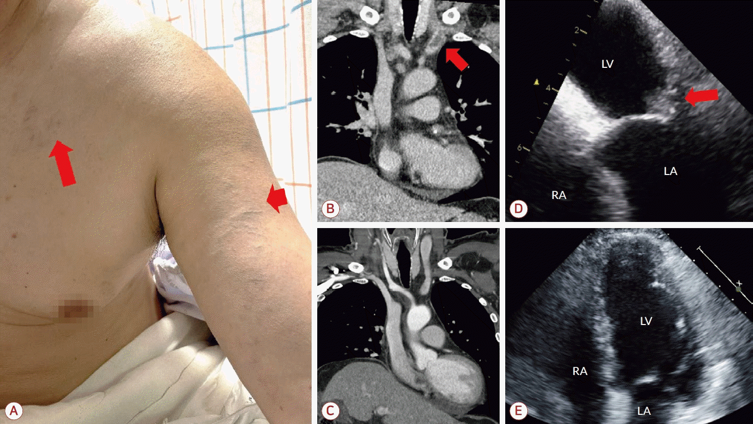
Skin lesion, thorax CT and echocardiography. (A) Superficial vein engorgements (arrows) are seen in left arm and subclavian area. (B) A thrombus at left brachiocephalic vein was detected on thorax CT (arrow). (C) Follow-up CT shows loss of thrombus in left brachiocephalic vein. (D) Trans-esophageal echocardiography shows a mass (0.44×0.45 cm) in the mitral valve (arrow). (E) Follow-up echocardiography shows loss of mass in the mitral valve. LV; left ventricle, LA; left atrium, RA; right atrium, RV; right ventricle, CT; computed tomography.
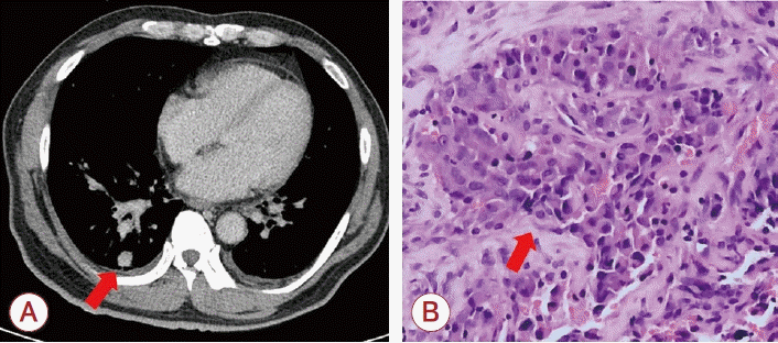
Lung CT and histopathologic findings of biopsy. (A) CT image shows a single nodule in right lower lung lobe (arrow). (B) Lymph node biopsy at left supraclavicular area with Hematoxylin-eosin staining (×400) shows poorly differentiated metastatic pulmonary adenocarcinoma (arrow). CT; computed tomography.
이후 환자는 항응고제(에독사반 60 mg) 일 1회 복용을 지속하였고, 입원 29일째 시행한 흉부경유심초음파 추적검사에서 승모판 부위 덩이는 확인되지 않았다(Fig. 2-E). 환자는 종양혈액내과에서 항암 치료를 시작하였고, 2개월 뒤 추적 흉부 CT에서 정맥 내 혈전은 소실되었으며(Fig. 2-C), 퇴원 후 3개월이 경과된 시점까지 뇌경색이나 정맥혈전증의 재발은 없었다.
고 찰
본 증례는 급성 허혈뇌졸중으로 내원한 환자에서 드문 부위에 반복되는 상지심부정맥혈전증과 비세균혈전심내막염의 원인으로 폐샘암종을 밝혀냈다.
심부정맥혈전증이 상지에서 발생하는 경우는 전체 심부정맥혈전증 환자들의 5% 정도로 매우 드물며, 대부분은 이차성 혈전으로 발생한다[4]. 액와 혹 은 쇄골하정맥혈전증의 경우, 어깨나 목 쪽의 애매한 불편감과 상지 부종 등을 호소하며, 피부정맥 확장, 목정맥(jugular vein) 확장, 촉지경성정맥(palpable venous cord) 등이 징후로 확인된다[1]. 본 증례에서도 왼쪽 가슴과 상지 쪽 피부정맥이 확장되어 있어 상지심부정맥혈전증을 의심할 수 있었다. 혈전증 및 지혈에 관한 국제과학표준화위원회(Scientific and Standardization Committee of the International Society on Thrombosis and Haemostasis)에서 제시하는 가이드라인에 따르면, 상지심부정맥혈전증과 같이 예기치 못한 부위에서 정맥혈전증이 발생한 환자는 잠재된 암의 가능성에 대해 여러 선별검사를 진행할 것을 권고하고 있다[5]. 상지심부정맥혈전증 환자에서 기본 치료는 최소 3개월 이상 항응고제를 사용하는 것이며, 증상의 정도 등에 따라 혈전용해 치료나 기계적혈전제거술 등을 시행할 수 있다. 본 증례에서는 저분자량헤파린 사용 중 폐샘암종이 확진된 후 심부정맥혈전증 및 뇌경색의 이차 예방을 위하여 최근 진행된 임상 연구에 근거하여 항응고제인 에독사반(edoxaban) 60 mg을 일 1회 투여하였다[6]. 그 결과 통 증 및 부종 등의 증상에서 호전을 보였으며, 이후 혈전이 없어짐을 확인하였다.
비세균혈전심내막염은 매우 드문 뇌경색의 원인 중 하나로, 항인지질항체증후군, 전신 홍반루푸스, 악성종양(malignancy), 응고항진성(hypercoagulability)과 관련되어 있으며, 심장 판막에 주로 혈소판으로 구성된 혈전이 침착되어 발생하는 것으로 알려져 있다[7]. 특히 암종의 경우, 종양괴사인자-1, 인터루킨-1과 같은 사이토카인 농도 상승이 혈관내피에 혈소판 응집을 잘 일으키며, 혈류가 빠른 심장 판막 부위에서 혈소판 응집이 잘 이루어지면서 발생하는 것으로 설명된다.[8]. 본 증례에서는 환자가 우측 팔머리정맥혈전증으로 이미 3개월 전부터 항응고제(rivaroxaban)를 복용하던 중에 뇌경색이 발생한 점에서, 항응고제에 잘 반응하지 않은 혈전이 원인이었을 가능성을 시사한다. 또한 이차 원인을 배제하기 위해 시행했던 자가면역 관련 검사에서도 모두 음성이었기 때문에 악성종양의 가능성을 두고 검사를 진행하였다.
입원 당시 가슴 단순X선검사에서 이상 병변을 시사하는 소견은 없었고 D-이합체 2,439 ng/mL가 확인되었으며, 심초음파검사를 통해 확인된 승모판 덩이로 Duke 진단기준에 따라 검사 및 치료를 진행하여 잠재된 악성종양에 대한 평가가 다소 늦어졌다. 따라서 예기치 못한 상지정맥혈전증과 비세균혈전심내막염 등 뇌경색 환자에게서 보기 드문 형태의 혈전증을 동반한다면 반드시 이차적인 원인에 대한 적극적인 검사가 필요하다는 것을 시사하는 증례로 문헌 고찰과 함께 보고한다.