심장 유두모양 섬유탄력종의 박리에 의한 급성뇌경색
Acute Ischemic Stroke Caused by Detachment of Cardiac Papillary Fibroelastomas
Article information
Trans Abstract
Cardiac papillary fibroelastomas are associated with a high risk of systemic embolization in spite of benign nature. We report a case of 85-year-old patient with left supraclinoid internal carotid artery occlusion who treated with aspiration thrombectomy. Despite of the absence of residual mass on echocardiography, we could analyze pathologic specimens using retrieved embolus and confirmed cardiac papillary fibroelastoma as a rare cause of stroke.
심장 유두모양 섬유탄력종(cardiac papillary fibroelastomas)은 원발심장종양 중에서 점액종 다음으로 많이 발견된다고 알려져 있으며, 부검 연구들에서 약 0.33%까지의 유병률이 보고되어 있다[1-4]. 또한, 심장 유두모양 섬유탄력종은 양성종양으로 무증상으로 발견되는 경우가 대부분이지만, 종양 자체의 탈락 또는 엽상체의 표면에서 형성된 섬유소-혈소판 응집에 의한 전신 색전증으로 인해 뇌경색을 포함한 다양한 합병증을 유발할 수 있다[5-10]. 저자들은 좌측 내경동맥 원위부 폐색으로 내원한 급성뇌경색 환자에서 기계적 혈전제거술을 통해 내경동맥을 재개통한 후 획득한 혈전의 병리 소견으로 뇌경색의 드문 원인인 심장 유두모양 섬유탄력종을 확인하여 보고하고자 한다.
증 례
85세 남자 환자가 2시간 전부터 발생한 우측 편마비 및 언어장애로 응급실에 왔다. 환자는 평소 흉통, 호흡곤란 및 실신 등의 증상 없이 지냈으며, 고혈압을 진단받고 혈압약과 아스피린을 복용 중이었다. 내원시 혈압은 175/69 mmHg, 맥박 58회였으며, 심전도는 정상동리듬이었다. 신경계진찰에서 의식은 혼미하였고, 완전실어증, 우측 중추안면마비, 우측 편마비(Medical Research Council [MRC] 1등급), 좌측 안구편위, 우측 동측반맹이 확인되었고 National Institute of Health 뇌졸중척도는 27점이었다.
비조영증강 뇌 computed tomography (CT)에서 뇌출혈의 소견이 없어 정맥내 재조합조직플라스미노겐활성제(intravenous recombinant tissue plasminogen activator)를 투여하였고, CT혈관조영술에서 좌측 내경동맥 원위부의 폐색이 확인되었다(Fig. 1). 흡인성도관을 이용한 기계적 혈전제거술을 시행하여, 약 3 cm의 점액질 양상의 혈전을 제거하여 modified thrombolysis in cerebral infarction 등급 2C의 재관류를 획득하였다(Figs. 2, 3-A). 혈전제거술 시행 후 24시간째 촬영한 뇌 CT에서 시술과 관련한 합병증은 보이지 않았으며, 신경계진찰에서 의식은 명료하였고 우측 상하지 근력 MRC 4등급으로 호전되었다.
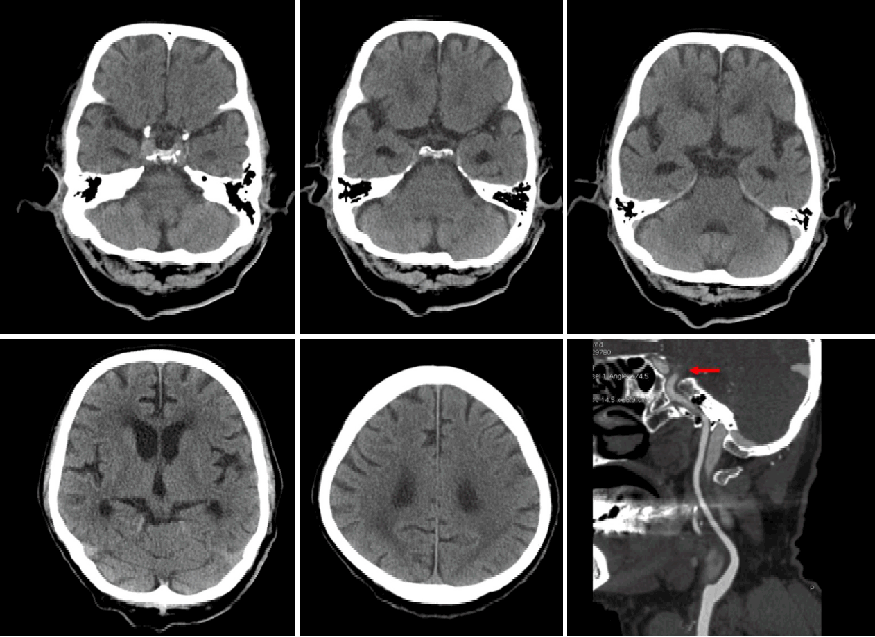
Baseline brain images of the patient. Brain computed tomography (CT) (A-E) and CT angiography (F, curved planar reformation image) showed occlusion of left supraclinoid internal carotid artery (arrow) without hyperdense vessel sign and evidence of acute ischemic stroke and hemorrhage.

Endovascular procedure of the patient. (A) Cerebral angiography showed left supraclinoid internal carotid artery occlusion (red arrow). (B) Mechanical thrombectomy was performed using aspiration catheter (white arrow). (C) Final angiography revealed modified Treatment In Cerebral Ischemia 2C of reperfusion.
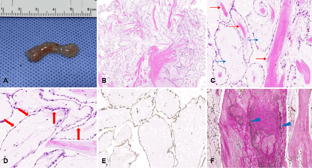
Pathologic features of the retrieved embolus. (A) Gross specimen from aspiration thrombectomy. (B) Embolus shows a papillary frond structure (H&E stain, ×20). (C) The frond is composed of avascular, paucicellular collagen bundles (red arrows) and myxoid matrix (blue arrows) (H&E stain, ×100). (D) The frond is lined with single layer of endothelial cells (arrows) (H&E stain, ×200). (E) The lining endothelial cells are positive for CD31 (CD31 immunohistochemical stain, ×200). (F) Elastic fibers are present in the central core of collagen (arrowheads) (Verhoeff’s elastic stain, ×100).
심인성 색전증 가능성을 고려하여 시행한 흉벽경유 심초음파검사에서 심장내 잔여 종괴 및 줄기(stalk)는 관찰되지 않았고 경한 승모판역류 외 이상 소견은 없었다. 식도경유 심초음파검사는 환자의 협조가 되지 않아 시행하지 못하였다. 혈관내 시술과정에서 획득한 혈전의 면역조직화학염색에서 심내막세포가 유두모양 엽상체를 단층으로 감싸고 있는 무혈관 유두종이 확인되었으며, 내부에는 탄력 섬유층이 확인되어 색전의 원인으로 심장 유두모양 섬유탄력종을 진단할 수 있었다(Fig. 3-B-F). 내원 당시 시행한 CT 혈관조영술 및 흉벽경유 심초음파검사 잔여 종괴는 확인되지 않았으나 작은 크기의 잔여 종괴 가능성을 배제할 수 없어 와파린 항응고 치료를 시작한 후 재활 치료를 위해 전원하였고, 퇴원시 NIH뇌졸중척도는 14점이었으며, 3개월 수정Rankin척도는 3점이었다.
고 찰
심장 유두모양 섬유탄력종은 과거에는 사후 부검에 의해 우연히 발견되는 경우가 많았으나, 현재는 심장 초음파 및 magnetic resonance imaging검사 등의 발달로 진단이 용이해졌으며 최근 심장 초음파를 이용한 연구들에서는 점액종보다 더 높은 빈도로 진단되기도 한다[3,4]. 심장 유두모양 섬유탄력종은 주로 대동맥판막 혹은 승모판막에서 발견되는 경우가 많으며, 보통 20 mm 이하의 작은 크기로 짧은 줄기(stalk)에 의해 심내막에 부착된 다발성 유두모양 엽상체(frond)모양을 하고 있다. 확진을 위해서는 병리조직 진단이 필수적이며, 한 층의 심내막세포가 탄력섬유를 포함한 무혈관의 결합조직 중심을 덮고 있는 점이 특징적이다. 본 증례에서는 이러한 특징적인 병리 소견이 확인되었으며, 흉벽경유 심초음파검사에서 심장내 잔여 종괴가 발견되지 않았지만 심장 유두모양 섬유탄력종으로 진단할 수 있었다.
심장 유두모양 섬유탄력종은 진단할 당시 무증상인 경우가 흔하지만, 증상을 동반하는 경우 호흡곤란(37.5%), 일과성허혈발작(37.5%), 흉통(12.5%), 실신(12.5%) 등이 보고되어 있다[4]. 해외의 후향적 연구에 의하면 심장 유두모양 섬유탄력종을 수술적으로 제거하지 않은 경우 진단 1년째 6%, 5년째 13%의 뇌졸중 발생률이 보고되었으며, 종양의 크기, 유동성 등의 심장 초음파 소견으로는 예후를 평가하는 데 충분하지 않아 수술 치료의 금기가 없는 경우 좌측 심장에 위치한 종양에 대해서 적극적인 절제를 권고하기도 하였다[3,4].
심장 유두모양 섬유탄력종에 의한 색전 뇌경색 환자들에서 급성기 재관류 치료로 정맥내 혈전용해제를 투여하거나 기계적 혈전제거술을 시행한 사례들이 보고되어 있다[5-10]. 혈전의 발생기전에 따라 엽상체의 표면에서 형성된 섬유소-혈소판 응집에 의한 색전증의 경우 혈전용해제가 효과적일 수 있으나[5,8], 심장 유두모양 섬유탄력종의 일부 또는 전체가 탈락되어 발생한 색전증의 경우 기계적혈전제거술이 보다 효과적일 것이다[8-10]. 본 증례는 병리 소견에서 섬유소-혈소판 응집 없이 유두모양 섬유탄력종이 확인되어 후자에 해당하는 경우로 생각되며, 흡인성 도관을 이용하여 부서지기 쉬운 점액질 양상의 색전을 보다 효과적으로 제거할 수 있었다.
저자들은 심장 유두모양 섬유탄력종에 의한 뇌경색 환자에서 기계적혈전제거술을 통해 효과적인 재관류를 하였으며, 가슴경유 심장 초음파검사에서 심장내 잔여 종괴가 없었지만 제거된 색전을 이용한 병리검사를 통해 뇌경색의 원인으로 심장 유두모양 탄력섬유종을 진단하여 이에 대해 보고한다.