주기적인 기관절개튜브 교체를 하는 환자에서 발견된 팔머리동맥박리
Brachiocephalic Trunk Dissection Found in Patient Undergoing Periodic Tracheostomy Tube Replacement
Article information
41세 남자가 발열 및 피(섞인)가래를 주소로 내원하였다. 17년 전 경부척수손상으로 사지마비가 발생하였고, 17년 전 기관절개술 후 주기적으로 기관절개튜브를 교체하고 있었다. 흉부 전산화단층촬영에서 폐렴 외에, 팔머리동맥에 충전결손이 발견되었다(Fig. 1). 뇌 자기공명영상 및 혈관조영술에서 팔머리동맥박리 및 우측 중대 뇌동맥 뇌경색이 관찰되었다(Fig. 2). 1년 전 흉부 전산화단층촬영에서 팔머리동맥의 충전결손은 없었으며, 13일 전에 기관절개튜브를 교체하였다. 충전결손이 기관절개튜브와 가까이 있었으므로 기관절개튜브 교체와 연관되어 팔머리동맥박리 및 뇌경색이 발생한 것으로 추정하였다.
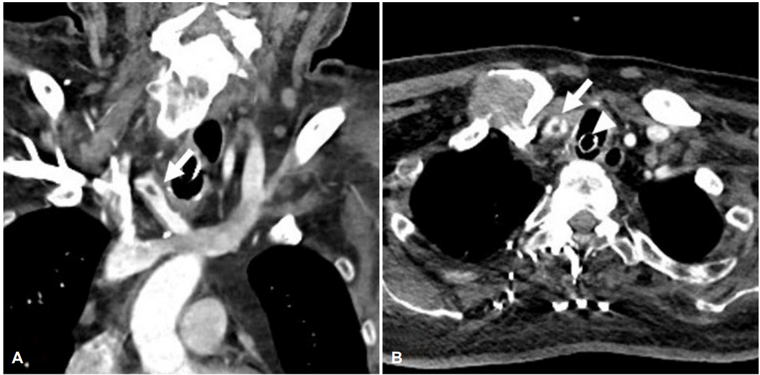
Enhanced chest CT image of the patient. (A) Chest CT coronal image reveals focal filling defect within the brachiocephalic trunk (white arrow). (B) Chest CT axial image reveals tear of brachiocephalic trunk arterial wall (white arrow) and tracheostomy tube (white arrowhead). CT; computed tomography.
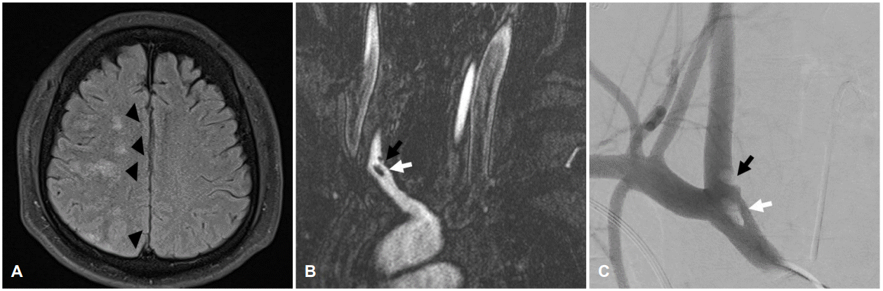
(A) FLAIR sequence of brain MR image shows multiple hyperintense foci compatible with subacute stage of embolic infarction (black arrowheads). (B) Contrast enhanced MR angiography and (C) conventional angiography image show focal filling defects in brachiocephalic trunk (white arrow) and proximal right CCA (black arrow), suggestive of dissection. FLAIR; fluid attenuated inversion recovery, MR; magnetic resonance, CCA; common carotid artery.
기관절개술 이후 팔머리동맥의 손상이 발생한 증례들은 보고되었으나, 기관절개튜브 교체 이후 팔머리동맥박리 및 뇌경색이 발생한 증례는 매우 드물다[1,2]. 기관절개튜브의 지속적인 교체가 요구되는 환자에서 팔머리동맥박리의 발생에 대한 주의가 필요하며, 영상검사에서 팔머리동맥의 충전결손이 발견될 경우 박리의 가능성 및 뇌경색에 대한 검사가 필요하다.