스쿠버다이빙이 원인으로 추정되는 내경동맥박리로 인한 앞맥락동맥 영역 뇌경색
Anterior Choroidal Artery Territory Infarction due to Internal Carotid Artery Dissection Presumably Caused by Scuba Diving
Article information
Trans Abstract
We report a case of anterior choroidal artery territory infarction due to internal carotid artery dissection presumably caused by scuba diving. A 44-year-old man presented with left facial palsy and hemiparesis. He had a history of scuba diving for 18 months. His last dive was 7 days ago, and he skipped decompression practice at that dive. We assumed that repetitive traumas and microbubbles during scuba diving, which made endothelium vulnerable to damage may have caused a carotid dissection.
스쿠버다이빙 이후 발생한 신경계합병증은, 일반적으로는 감압병과 그에 따른 공기색전증으로 인한 허혈 증상이 알려져 있다. 이 경우, 고압산소 치료를 통해 회복이 가능하다[1]. 한편 스쿠버다이빙 이후 발생한 내경동맥박리 및 이와 동반된 뇌경색증에 대한 보고가 증가하고 있으나[2-7], 국내에서의 보고는 현재까지 확인되지 않았다. 저자들은 스쿠버다이빙 이후 발생한 것으로 추정되는 내경동맥박리 환자를 경험하여, 이를 보고하고자 한다.
증 례
44세 남자가 내원 4시간 전 갑자기 발생한 왼쪽 안면 및 상지마비로 타 병원의 응급실을 방문하였고, 뇌 영상검사 시행 후 상급병원으로의 전원을 권유받아, 본원으로 전원되었다. 우측 측두부 및 안구 주위 통증이 상지 마비와 동시에 생긴 뒤 즉시 호전되었으며, 안면마비는 4시간 뒤 호전되었다. 당뇨병, 고혈압, 흡연 등의 혈관위험인자는 확인되지 않았다. 신경계진찰에서, 좌측 상지 근력은 Medical Research Council 4등급이었고, 우측 호르너증후군(안검하수 및 부분 안면 무한증)이 관찰되었다. 환자는 18개월 전 스쿠버다이빙을 시작하여 매달 2-3회 정도 하였으며, 내원 7일 전 마지막 스쿠버다이빙을 하였다. 당시, 수심 15미터까지 잠수한 뒤 감압과정을 생략하였고, 환자는 수면에 올라온 직후 약한 어지럼증을 호소하다가 호전되었다.
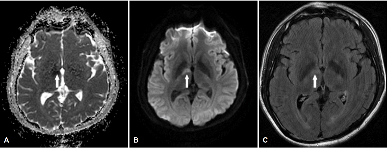
내원 당일 시행한 뇌 확산강조영상 및 겉보기확산계수지도에서 우측 앞맥락동맥 영역의 고신호 및 저신호강도가 관찰되었으며, 액체감쇠역전회복영상에서도 동일 영역의 고신호강도가 확인되었다(Fig. 1). 대퇴동맥경유뇌혈관조영술(transfemoral cerebral angiography)에서 우측 두개외내경동맥의 ‘불꽃모양 폐색(flame-shaped occlusion)’이 확인되었으며, 동맥박리에 합당한 소견으로 판단되었다. 우측 전대뇌동맥 및 중대뇌동맥 혈류는 전교통동맥을 통해 좌측으로부터 공급되었다(Fig. 2). 혈액검사에서 혈구수치, 간 및 신장기능, 고지혈증검사 등은 정상이었고, 심장 초음파검사에서 특이 소견은 없었다. 경동맥박리의 선행요인으로 알려진 마르팡증후군, 섬유근형성이상(fibromuscular dysplasia), Ehlers-Danlos증후군 등과 관련한 임상이나 영상 소견이 없어, 추가검사는 시행하지 않았다. 항핵항체, 항호중구세포질항체(anti-neutrophil cytoplasmic antibody, ANCA), 항SSA 및 SSB항체, 항카디오리핀항체, 항 이중나선DNA항체는 모두 음성이었다. 혈액응고인자검사에서 C단백질 및 S단백질, 피브리노겐 등은 모두 정상이었다.

Axial view of magnetic resonance image shows hypointensities on apparent diffusion coefficient (ADC) map in the right genu and posterior limb of internal capsule (white arrow, A), diffusion restriction (white arrow, B), and mild hyperintensities on fluid-attenuated inversion recovery (FLAIR) image at the same lesion (white arrow, C), suggesting acute infarction of the right anterior choroidal artery territory.

A lateral view from right common carotid angiogram (A) demonstrates flame-shaped occlusion of the right proximal internal carotid artery, consistent with acute dissection. A Town's view from left carotid angiogram (B) shows good cross-filling through the anterior communicating artery.
폐색된 내경동맥의 재개통 및 뇌경색의 2차예방 목적으로 항응고제(와파린 3.5 mg) 및 항혈소판제(아스피린 100 mg) 복합요법을 시작하였고, 프로트롬빈시간 국제정상화비율(prothrombin time international normalized ratio, PT INR) 1.84를 확인한 뒤, 항혈소판제는 중단하였다. 환자는 10일의 입원 기간 동안 신경계 증상은 모두 호전되었으며, 3개월 후 시행한 추적 컴퓨터단층혈관조영에서 내경동맥 폐색이 지속되어 PT INR 2-3 유지를 목표로 항응고제 치료를 지속하였다.
고 찰
본 증례는 지속적인 스쿠버다이빙의 병력, 영상검사와 임상 양상에서 확인된 내경동맥박리, 젊은 나이의 남성 및 다른 혈관위험인자가 없는 점, 유사한 병력의 내경동맥박리 환자들의 보고가 있음을 고려하여, 스쿠버다이빙과 연관성이 있는 내경동맥박리로 판단된다.
스쿠버다이빙 후 경동맥박리가 발생하는 기전으로, 입수 및 상승, 하강 등의 과정 중 발생하는 과도한 경부신장 및 경부회전에 따른 동맥 내막의 손상 그리고 차가운 물에 입수할 때 교감신경이 활성화되면서 나타나는 혈압 상승 등이 주요 원인으로 알려져 있다[4,6]. 운동 중 발생하는 경부혈관박리는 운동 종목에 따른 차이가 알려져 있고, 스쿠버다이빙의 경우 전방순환계(anterior circulation) 뇌졸중의 발생 비율이 후방순환계에 비해 높다[8]. 다이빙 경험 정도에 따른 비교 결과에서는 초심자부터 숙련된 다이버까지 다양하게 경부혈관박리가 보고되었다[2]. 내경동맥박리는 대부분 경부 혹은 두부 통증과 함께 뇌허혈 증상을 동반하는 경우가 많으며, 허혈 증상을 유발하지 않는 박리증의 경우, 호르너증후군이나 아래뇌신경마비(lower cranial nerve palsy)를 잘 동반한다[5]. 발생 시기는 대개 운동 중에서부터 1일까지 가장 많은 편이나, 2일 뒤에 발생한 사례도 있다[9].
본 증례는 운동 1주일 뒤 뇌경색이 발생하였지만, 반복적인 스쿠버다이빙에 따른 외상으로 인한 내경동맥 내막의 손상이 박리 발생에 영향을 준 것으로 추정된다. 또한, 스쿠버다이빙 활동에서 감압과정 없이 수면에 빠르게 도달할 경우, 혈액 속 질소분자가 미세공기방울(microbubbles)로 변하는 현상이 보고되고 있으며, 이 공기방울 및 그와 연관된 혈관내 물질들이 내막손상을 일으킨다고 보고되었다[10]. 본 증례에서도 미세공기방울의 영향이 있을 것으로 추측된다.
최근 생활수준의 향상으로 국내에서도 스쿠버다이빙을 즐기는 인구가 증가할 것이므로, 이에 따라 내경동맥박리의 발생 가능성도 늘어날 것으로 판단한다. 스쿠버다이빙의 병력이 있는 신경계 이상 소견의 환자를 진료할 때, 감압병뿐만 아니라, 내경동맥박리 가능성 또한 고려해봐야 하겠다.