

50세 남자가 최근 한 달 전부터 가끔씩 발생하는 두통으로 내원하였다. 두통은 긴장형두통 양상으로 35년 전 두개내 출혈의 과거력이 있어 두부 컴퓨터단층촬영을 시행하였고, 양측에 거대한 석회화를 동반한 병변과 이로 인한 뇌의 위축이 보였으며 뇌 자기공명영상를 통해 석회화된 만성경막하혈종으로 진단하였다(Fig.). 이는 만성경막하혈종의 0.3-2.7%에서만 나타날 정도로 매우 드문 후유증으로, 임상 증상은 정신지체, 치매, 국소 신경계 징후 등 다양하게 나타나는 것으로 알려져 있지만 본 증례와 같이 드물게 증상이 없을 수도 있다[1]. 증례에 따라 수술을 하는 경우도 있으나 본 증례의 경우는 일반 진통제에 반응이 좋아 수술을 하지 않았다[2].
| J Korean Neurol Assoc > Volume 37(2); 2019 > Article |

|
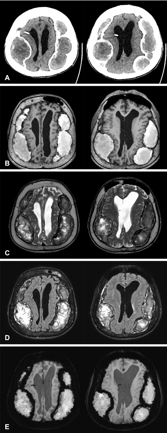
Figure.
Brain computed tomography shown multiple extra-axial crescent-shaped or bi-convex-shaped hypodense mass like lesions with thick hyperdense calcific margins (A). Brain magnetic resonance images (MRI) shown huge hyperintense lesions to cerebrospinal fluid (CSF), with hypointense capsule to CSF on T1 images (B). On T2 images shown the lesions that isointense to CSF with hypointense capsule (C). The lesions were hyperintense and the capsule was hypointense compare with CSF intensity on fluid attenuated invention recovery images (D). Susceptibility weighted images shown markedly hypointense periphery (E).
- TOOLS
 PDF Links
 PubReader
 ePub Link
 Full text via DOI
 Download Citation
 Print
Print


-
METRICS

-
- 0 Crossref
- 0 Scopus
- 3,078 View
- 64 Download
-

- Related articles
-
An Autopsy Case of Amyotrophic Lateral Sclerosis with Neuroinflammatory change2012 ;30(2)
A Case of Cryptococcal Meningitis Presenting as Bilateral Sensorineural Hearing Loss2006 ;24(3)
A Case of Cerebral Infarction and Chronic Subdural Hematoma in Essential Thrombocythemia2000 ;18(2)
A Case of Bilateral Cavernous Sinus Mucormycosis1996 ;14(3)
A Case of Dern atomyositis Associated with Primary Hepatoma1989 ;7(1)
- Editorial Office
-
(ZIP 03163) #1111, Daeil Bldg, 12, Insadong-gil, Jongno-gu, Seoul, Korea
Tel: +82-2-737-6530 Fax: +82-2-737-6531 E-mail: jkna@neuro.or.kr
Copyright © 2024 by Korean Neurological Association.

