

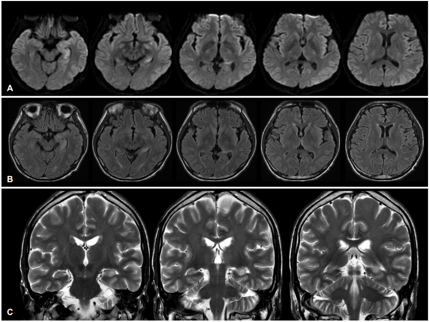
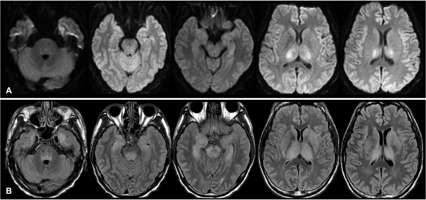
기저질환이 없는 47세 남자가 하루 전 발생한 열로 본원 응급실에 왔다. 체온은 38.5℃였고, 신경학적 진찰시 의식상태는 졸린 상태였다. 뇌척수액검사에서 색은 투명하였고, 압력은 70 mmH2O였으며, 적혈구 3/μL, 백혈구 12/μL, 단백질 51 mg/dL, 포도당 88 mg/dL로 확인되었다. 혈청 및 뇌척수액에서 단순헤르페스바이러스검사는 음성이었다. 뇌 자기공명영상에서 좌측 해마의 몸통과 꼬리에 저명한 고신호강도가 관찰되었고, 그 외 양측 내측 시상 병터가 의심되었다(Fig. 1). 조영증강 뇌 자기공명영상에서 특이 소견은 보이지 않았다. 2일 후 환자는 혼수상태로 악화되었고, 뇌간반사가 소실되었다. 이후 혈청에서 시행한 효소결합면역흡착측정법(enzyme-linked immunosorbent assay)에서 일본뇌염바이러스 특이 immunoglobulin M 항체가 확인되었다. 2주 뒤 추적 뇌 자기공명영상에서 뇌간, 시상, 기저핵, 대뇌피질에서 광범위한 병터가 관찰되었다(Fig. 2).
| J Korean Neurol Assoc > Volume 37(1); 2019 > Article |

|
REFERENCES
1. Handique SK, Das RR, Barman K, Medhi N, Saharia B, Saikia P, et al. Temporal lobe involvement in Japanese encephalitis: problems in differential diagnosis. AJNR Am J Neuroradiol 2006;27:1027-1031.
2. Linn J, Danek A, Seelos KC, Brückmann H. Differential diagnosis of bilateral thalamic lesions. Clin Neuroradiol 2007;17:3-22.

- TOOLS
 PDF Links
 PubReader
 ePub Link
 Full text via DOI
 Download Citation
 Print
Print


-
METRICS

-
- 0 Crossref
- 0 Scopus
- 3,721 View
- 71 Download
-

- Related articles
-
Central Skull Base Osteomyelitis Presenting with Bilateral Abducens Nerve Palsy2021 November;39(4)
Herpes Simplex Encephalitis Presenting as An Acute Motor Aphasia2020 May;38(2)
Scrub Typhus Encephalitis Presenting with Unilateral, Multifocal Brain Lesions2020 February;38(1)
Meningeal Carcinomatosis Presenting with Isolated Pseudotumor Cerebri2020 February;38(1)
- Editorial Office
-
(ZIP 03163) #1111, Daeil Bldg, 12, Insadong-gil, Jongno-gu, Seoul, Korea
Tel: +82-2-737-6530 Fax: +82-2-737-6531 E-mail: jkna@neuro.or.kr
Copyright © 2024 by Korean Neurological Association.

