

| J Korean Neurol Assoc > Volume 36(4); 2018 > Article |

|
REFERENCES
1. Burnett MM, Hess CP, Roberts JP, Bass NM, Douglas VC, Josephson SA. Presentation of reversible posterior leukoencephalopathy syndrome in patients on calcineurin inhibitors. Clinl Neurol Neurosurg 2010;112:886-891.

2. Luu ST, Lee AW, Chen CS. Bilateral occipital lobe infarction with altitudinal field loss following radiofrequency cardiac catheter ablation. BMC Cardiovasc Disord 2010;10:14.
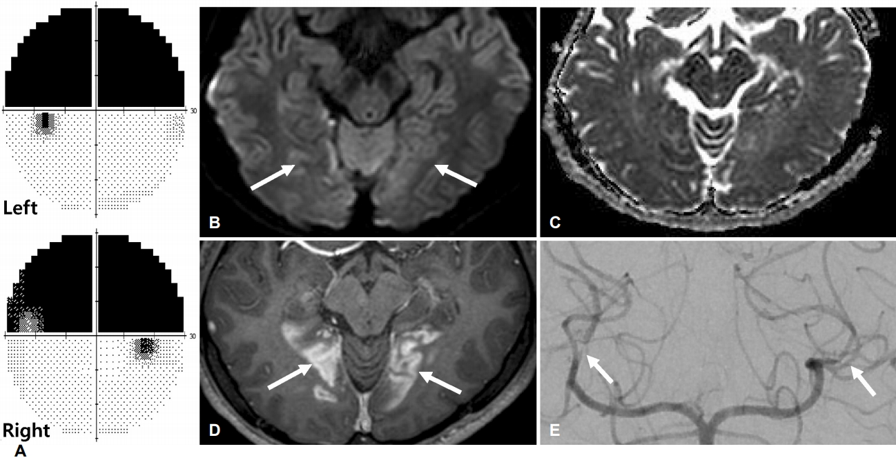
Figure.
(A) Automated perimetry using Humphrey program of the right and left eye showed bilateral superior altitudinal field defects. Brain magnetic resonance imaging scan axial view, (B) diffusion weighted image, (C) apparent diffusion coefficient image at corresponding level, and (D) T1 enhanced sequence, showing area of abnormal signal in both occipital lobes and extending into the lower bank of calcarine sulcus (white arrows), consistent with subacute cerebral infarction. (E) In cerebral angiography, focal filling defect (white arrows) suggesting vasospasm in both posterior cerebral artery was identified.
- TOOLS
 PDF Links
 PubReader
 ePub Link
 Full text via DOI
 Download Citation
 Print
Print


-
METRICS

-
- 0 Crossref
- 0 Scopus
- 3,483 View
- 69 Download
-

- Related articles
-
Posterior Reversible Encephalopathy Syndrome2016 November;34(4)
Hypertension-induced Posterior Reversible Encephalopathy Syndrome2001 ;19(5)
- Editorial Office
-
(ZIP 03163) #1111, Daeil Bldg, 12, Insadong-gil, Jongno-gu, Seoul, Korea
Tel: +82-2-737-6530 Fax: +82-2-737-6531 E-mail: jkna@neuro.or.kr
Copyright © 2024 by Korean Neurological Association.

