단독 혀마비로 발현한 항체 양성 시신경척수염범주질환
Isolated Tongue Paralysis as Presentation of Seropositive Neuromyelitis Optica Spectrum Disorder
Article information
Trans Abstract
Neuromyelitis optica spectrum disorder (NMOSD) is characterized by a characteristic clinical presentation or positivity for the anti-aquaporin-4 antibody. Lesions involving the dorsal medulla are typical of NMOSD, but isolated tongue paralysis has not been reported previously. We report a rare case of NMOSD presenting with isolated tongue paralysis and swelling due to intrinsic tongue muscle paralysis, which was caused by bilateral involvement of the hypoglossal nuclei in the lower dorsal medulla oblongata.
시신경척수염범주질환(neuromyelitis optica spectrum disorder, NMOSD)은 시신경염, 광범위한 척수염(extensive transverse myelitis), 맨아래구역(area postrema) 병변이 반복적으로 나타나거나, 특징적인 병변과 함께 anti-aquaporin 4 (AQP-4) 항체가 양성인 경우 진단할 수 있다[1]. NMOSD는 주로 뇌간과 연수부위를 침범하여 다양한 뇌간증상이 나타날 수 있지만, 초기에 뇌증상을 보이는 경우는 비교적 드물다[2]. 또한 다른 신경계증상의 동반 없이 뇌간증상 단독으로 나타나는 경우는 매우 드물며, 특히 단독 혀마비 또는 양쪽 설하신경마비를 보고한 적이 없다. 저자들은 등쪽 연수부위를 침범하여 단독 혀마비로 발현한 NMOSD 1예를 보고하고자 한다.
증 례
55세 여자가 한 달 전부터 나타난 삼킴곤란과 혀부종 때문에 응급실에 왔다. 환자는 삼킴 곤란과 함께 오심과 구토가 동반되어 2주 동안 아무 것도 먹지 못한 상태였다. 과거 병력이 없었고, 병원에 왔을 때 활력징후는 안정적이었다. 신체검사에서 혀는 양쪽이 모두 부풀어 커져 있는 상태였고, 표면에 염증이나 궤양은 없었다. 신경계진찰에서 의식은 명료하였고, 의사소통은 정상이었지만 설음을 발음할 때 심한 구음장애와 양측 설하신경마비가 있어서 혀내밀기는 불가능하였고, 좌우로 전혀 움직이지 못하는 상태였다. 구역반사는 정상적으로 나타났고, 나머지 뇌신경기능도 정상이었다. 사지 마비나 감각이상 또는 소뇌이상징후는 없었다.
응급실에서 촬영한 뇌자기공명영상(magnetic resonance imaging, MRI)의 확산강조영상과 T2 강조영상에서 연수 피개(tegmentum)와 양쪽 소뇌 치아핵(dentate nucleus), 좌측 후두엽의 뇌실주변백질에 고음영이 보였다(Fig. 1). 척수 MRI 그리고 시각유발전위, 청각유발전위와 체성감각유발전위검사는 정상이었다. Redmond 방법을 이용하여 설하신경전도검사를 하였고, Redmond 방법의 정상값을 기준으로 이상 여부를 판단하였다[3]. 혀를 잘 말린 후 한 쪽 혀의 등쪽에 금속 설압자를 놓고 그 위에 기록전극(recording electrode)과 기준전극(reference electrode)을 2 cm 간격을 두고 부착하고, 접지전극은 뺨에 부착하였다. 전기자극은 하악골의 아랫면을 따라 최대초과자극(supramaximal stimulation)이 될 때까지 서서히 올려 기록하였다. 왼쪽은 전기자극에 대해 반응이 없었고, 우측은 말단 잠복기가 5.5 ms로 지연되어 있었다 (참고치 2.2±0.4 ms). 혀근육의 침근전도검사에서 탈신경전위는 나타나지 않았고 운동단위의 동원이 감소되었다.
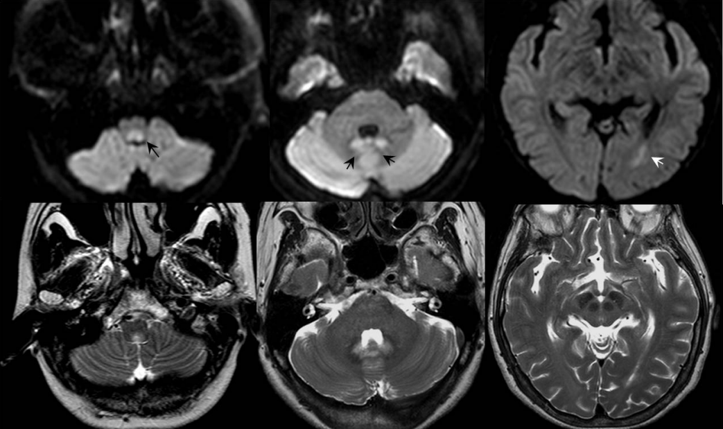
Brain MRI performed at first visit. Subtle T2-high and DWI-high signal intensity lesions were observed in tegmentum of medulla oblongata, both side cerebellum, and periventricular white matter of left occipital lobe (black and white arrows). MRI; magnetic resonance imaging, DWI; diffusion weighted imaging.
뇌척수액검사에서 압력 60 mmH2O, 백혈구 60/mm2 (mononuclear form 90%), 단백질 36 mg/dL, 당 62 mg/dL(혈당 91 mg/dL)였다. 면역글로불린G지수(IgG index)는 0.53으로 정상이었고, 올리고클론띠(oligoclonal band)는 보이지 않았다. 세균, 진균에 대한 염색 또는 배양검사와 바이러스항체검사는 정상이었다. 혈액검사에서 항SSA/Ro, 항SSB/La항체가 양성이었고, 항핵항체(anti-nuclear antibodies)검사도 양성이고 형광 패턴은 speckled형이었다. 쇠그렌증후군 진단을 위해 쉬르머검사와 아랫입술 부위에서 침샘조직검사를 하였으나 정상이었다. 혈장과 뇌척수액의 AQP-4항체는 양성이었다.
NMOSD로 진단하고 고용량 메틸프레드니솔론 정주요법(methylprednisolone pulse therapy, 1,000 mg/day for 5 days)을 시작하였고, 삼킴곤란으로 음식물 섭취가 불가능하여 비위관을 삽입하였다. 입원 3일째 다시 촬영한 뇌 MRI에서 처음 보였던 고음영이 더욱 뚜렷하게 나타났고 조영증강되었다(Fig. 2). 구인두 MRI에서 혀 내부에 침윤이나 염증병변은 없었고, 혀의 횡경선이 증가되어 있었다(Fig. 3).
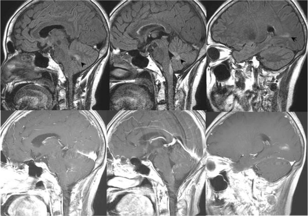
FLAIR and T1-enhanced MRI performed on 3 days after initial MRI. High signal intensity with contrast enhancement was shown in tegmentum of medulla oblongata (white arrow), both cerebellum (black arrowhead), and periventricular white matter (black arrow) of left occipital lobe. FLAIR; fluid attenuated inversion recovery, MRI; magnetic resonance imaging.

Oropharyngeal MRI. The transverse diameter of tongue base was enlarged on T2-weighted image (A), and there was no inflammatory or infiltrative disease in the tongue on T1-enhanced image (B). MRI; magnetic resonance imaging.
환자의 증상은 고용량 스테로이드치료로 호전되지 않았으나 증상이 악화되지 않고 다른 신경계징후가 새로 나타나지 않아, 경구용 스테로이드와 아자티오프린(azathioprine)으로 면역 치료를 유지하였고, 재활 치료를 통해 서서히 증상이 호전되었다. 구음장애와 삼킴 곤란은 서서히 호전되어 발병 1개월 후에 비위관을 제거하고 구강섭취가 가능하였고, 지금까지 2년 동안 외래에서 추적 중이며 증상 재발은 없었다.
고 찰
NMOSD는 anti-AQP-4항체가 양성이거나, 주로 시신경, 척수, 맨아래구역, 간뇌를 반복적으로 침범하는 병변에 의해 나타나는 임상증후군이다[1]. NMOSD의 뇌 MRI에서 병변을 보이는 경우는 43-70% 정도이지만[4], 첫 발현 증상이 뇌증상인 경우는 약 18-24%로 비교적 드물다[2].
NMOSD의 특징적인 뇌병변 위치는 AQP-4 발현이 높은 제3뇌실과 뇌수도관(cerebral aqueduct)을 둘러 싼 간뇌부위로 시상, 시상하부, 중뇌의 앞쪽 경계부위이며, 제4뇌실과 인접한 등쪽 뇌간인 맨아래구역과 고립로핵(nucleus tractus solitarius) 도 자주 침범한다[4]. NMOSD의 특징적인 병변 위치는 아니지만 측뇌실주변의 뇌량, 대뇌백질, 피질척수로 병변도 보고된 적이 있다[4]. 본 증례는 등쪽 연수, 소뇌 치아핵, 좌측 후두엽의 백질이 침범되었고, 이는 NMOSD에서 자주 침범되는 부위이다.
그러나 본 증례는 등쪽 연수에 비교적 넓게 차지하는 병변 때문에 양쪽 설하신경핵이 침범되어 전체 혀마비와 혀의 비대로 발현되었다. NMOSD에서 제3뇌실주변의 간뇌를 침범하는 경우, 항이뇨호르몬 분비이상, 기면증, 저체온증, 저혈압 같은 증세가 나타날 수 있고, 등쪽 뇌간을 침범하는 경우에는 난치성 딸국질, 오심, 구토가 나타난다. 또 안진, 구음장애, 삼킴곤란, 실조증, 안근마비 같은 다양한 뇌간증상이 생길 수 있으나, 단독 뇌간 증상이 나타나는 경우는 드물다. 다른 증례에서 양쪽 설하신경마비 또는 전체 혀마비가 연수경색[5]과 전이암[6]에 의한 설하신경의 압박에 의해 나타났다는 보고가 있으나, 아직 NMOSD에서 전체 혀마비가 주 증상으로 발현된 예는 없었다. 본 증례는 구역, 구토 증상을 동반하였으나, 신경계 이상은 혀마비가 유일하였고, 이 환자에서 보였던 혀의 부종은 양측 설하신경마비로 인한 내재근 마비와 이로 인한 근육긴장도의 감소로 인해 나타난 것으로 보인다[7].
본 증례는 항ENA항체 양성이었으나, 쇠그렌증후군의 임상징후는 없었다. Anti-AQP-4항체 양성인 환자에서 자가항체가 같이 발견되는 경우는 38-75% 정도이며[8], 이 중 anti-SSA/Ro항체가 흔하다. 그러나 재발성 척수염과 NMO로 진단받은 경우, 단발성 질환 보다 anti-SSA/Ro항체 양성이 더 많고[9], 쇠그렌증후군 환자 중에서 anti-AQP-4항체 양성인 경우에 NMO의 발병과 재발이 더 높은 것으로 알려져 있다[10]. 이는 쇠그렌증후군 같은 자가면역질환에서 반복적인 뇌신경계 침범이 있을 때, anti-AQP-4항체 확인이 필요하다는 것을 시사한다. 또 본 환자처럼 anti-AQP-4항체 양성이면서 다른 자가항체가 같이 발견되는 점은 NMO나 NMOSD도 다발성자가면역증후군(multiple autoimmune disease)의 범주에 속할 수 있음을 시사한다.
본 증례는 등쪽 연수를 주로 침범하는 특징적인 뇌병변과 anti-AQP-4항체와 SS-A/SS-B항체가 양성이어서 NMOSD로 진단하였고, 양측 설하신경병변으로 인해 다른 신경계 이상 없이 단독 혀마비가 주 증상인 드문 예이다.